A choroidal hemangioma is a benign vascular tumor of the choroid. It can be seen in syndromic pathology as a diffuse lesion and isolated as a primary ocular disease in the circumscribed variant.
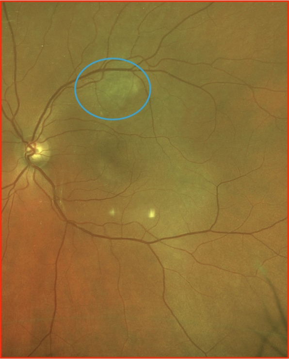
The following images represent a single case demonstrating the appearance of a choroidal hemangioma using multimodal imaging.
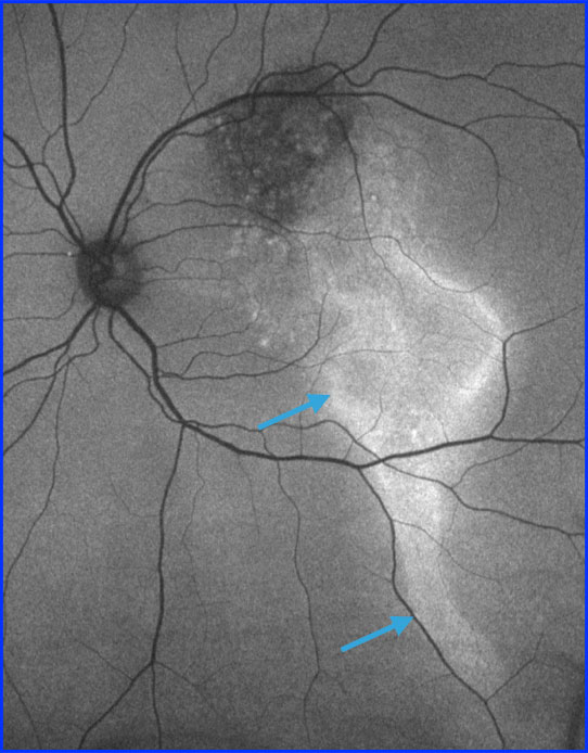
On autofluorescence the appearance of a gutter-like change (arrows) can be noted due to chronic fluid exudation in a gravity-dependent manner.
This OCT image is through the foveal center demonstrating subretinal fluid. This image is a useful reminder as to the importance of a good differential diagnosis for subretinal fluid.
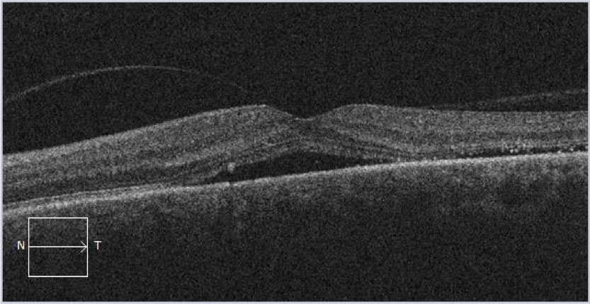
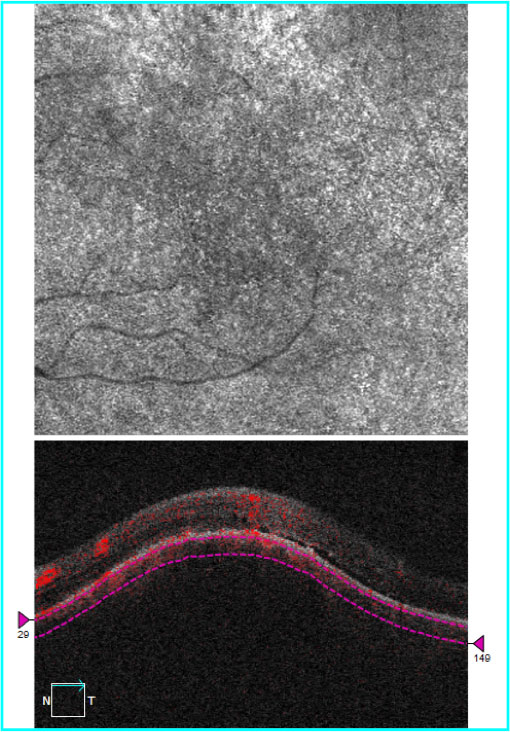
The OCT image through the lesion demonstrates bowing of the RPE over the underlying choroidal mass. A small amount of subretinal fluid is also noted. The OCT-Angiogram demonstrates some increased vascularity but in isolation could not be used for diagnostic confirmation.
A video file demonstrating the appearance of the lesion on a macular cube scan. Note that the superior aspect captures a small portion of the lesion and the remainder demonstrates subretinal fluid and atrophy inferiorly.
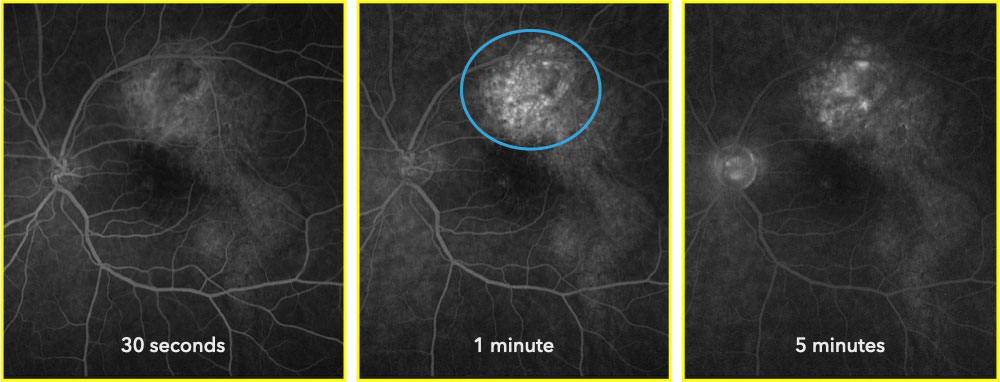
Fluorescein angiogram demonstrating stippled hyperfluorescence over the main aspects of the lesion (blue circle) with late leakage.