A coloboma is an area devoid of retinal and choroidal tissue.
Example 1: A coloboma noted inferiorly.

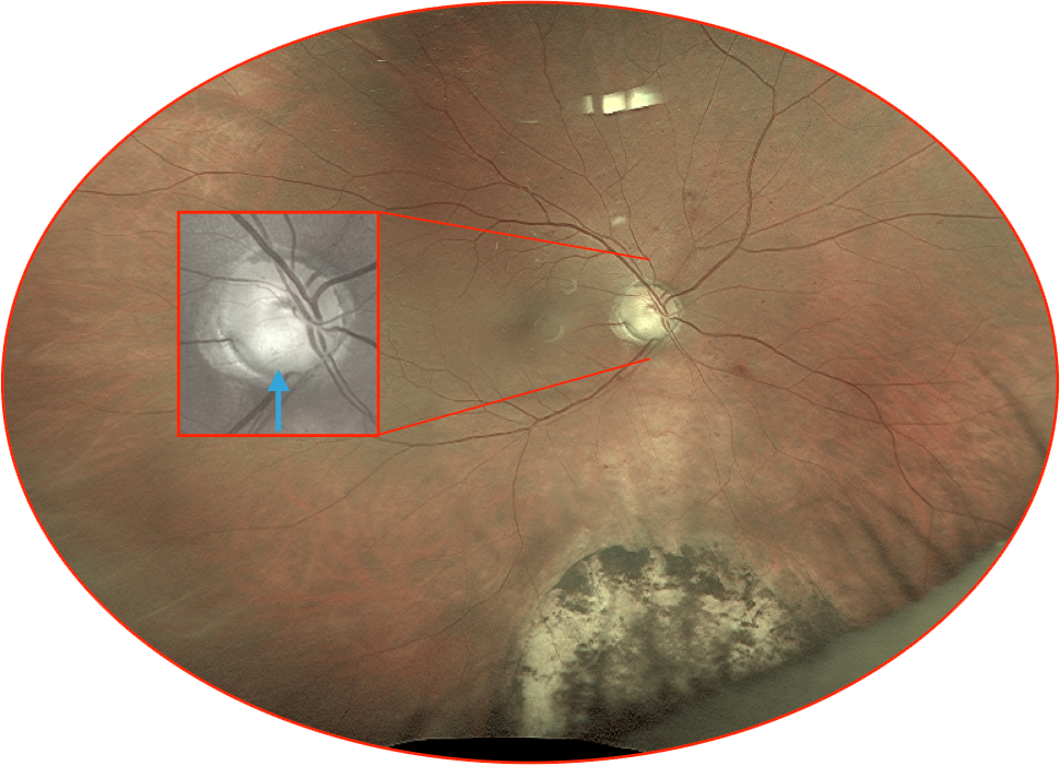
Example 2: There is an inferior coloboma and an accompanying coloboma of the optic nerve head (the color has been optimized for visualization and enlarged in the inset, blue arrow). This patient had an additional coloboma of the inferior iris (not shown).
Example 3, Part 1: A coloboma with corresponding autofluorescent imaging. While this is not in a classic location, the next image of the accompanying OCT of the overlying region is demonstratively useful.
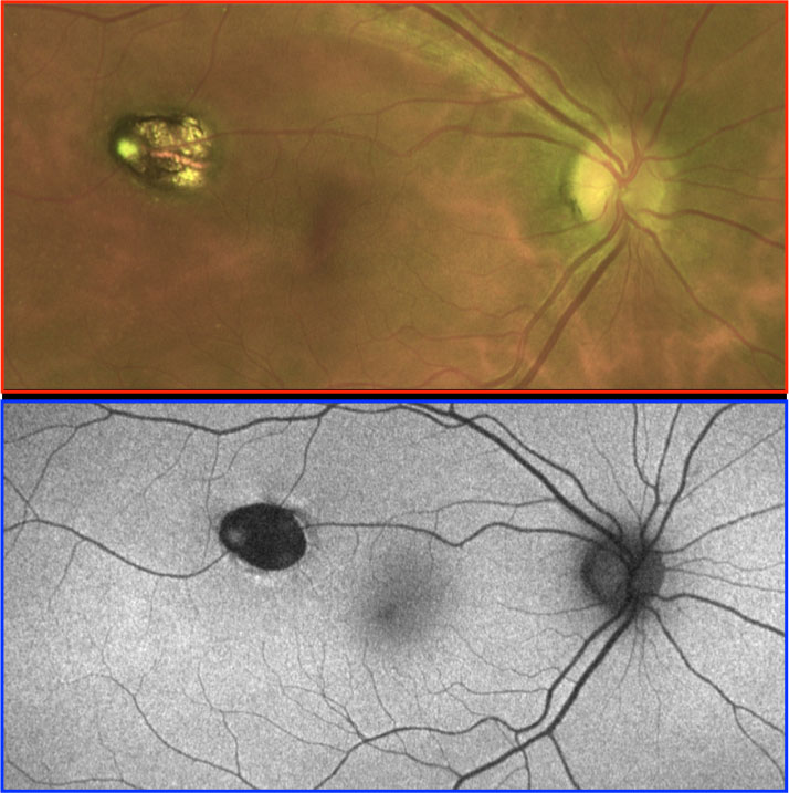
Example 3, Part 2: While not perfectly centered over the colobomatous lesion, this OCT gives the general idea as to the anatomy of a coloboma in-vivo. Note the profound loss of both retinal and choroidal tissue.