From our June 13, 2016 newsletter.
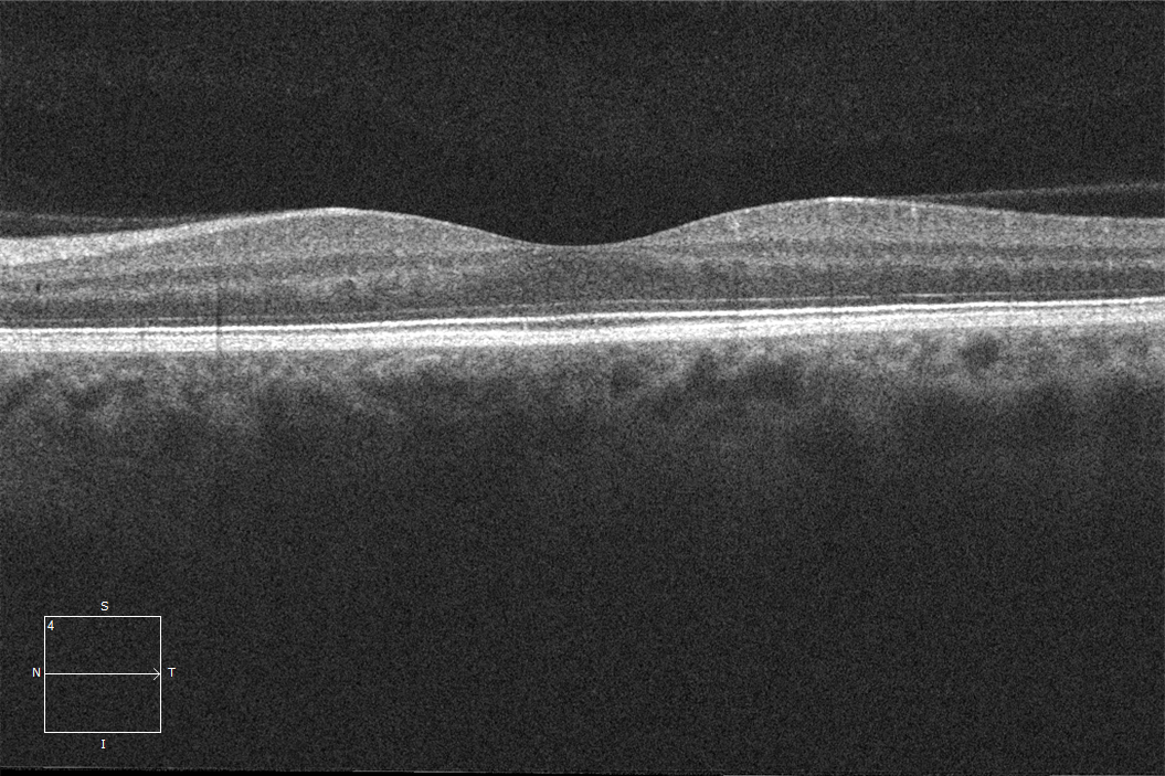
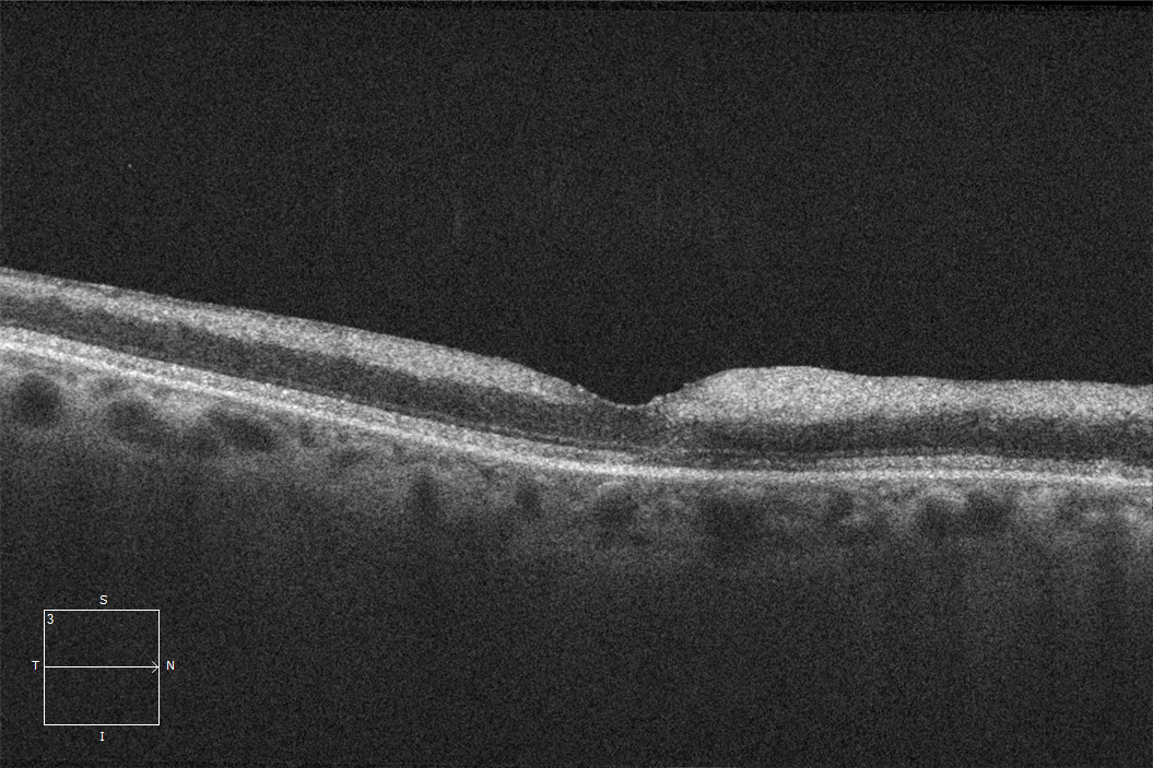
Inner retinal opacification is a common finding seen on OCTs for retinal artery occlusions. That’s because it shares the same pathophysiology as a cherry-red spot. Keep in mind that the inner retina is nourished by the retinal vasculature, while the outer retina is nourished by the choroidal vasculature. So… when you have a retinal arterial occlusion, it’s the inner retina that’s affected. The normally transparent retina loses its homeostatic mechanisms and opacifies. On OCT, this appears as both a thickening and an opacification.Tip: Hone your ability to detect subtle abnormalities by viewing as many images of normal retinas as possible.
When it comes to detecting subtle abnormalities, having a point of reference is so important. That’s why we recommend familiarizing yourself with as many normal images as you can get your hands on. Below is a side-by-side comparison of a normal retina and a retina with inner opacification and thickening, consistent with a central retinal artery occlusion. Would you have been able to identify the differences?A Clear Vision for Professional Development
Elevate your eye care skills with image-rich content and relevant case examples that empower you to treat patients more effectively.
Send this Newsletter to a Friend
