From our May 30, 2016 newsletter.
Now that we’ve gone over the difference between subretinal lucency and subretinal fluid in the last newsletter, we’d like to point out that the same concept can apply to intraretinal changes. The OCT lexicon continues to evolve and there are many unanswered questions, such as… Are all retinal cystic spaces filled with fluid? Or, do some maybe represent degenerative aspects of the nerves? In this case, the term “cyst” or “intraretinal fluid” may mean different things to different doctors.Here’s an example in cystoid macular edema
In cystoid macular edema, after a retinal vein occlusion, intraretinal fluid is common. The leakage of fluid from the damaged retinal vasculature leaves little doubt as to what is in the retina (i.e. it is extravasated fluid).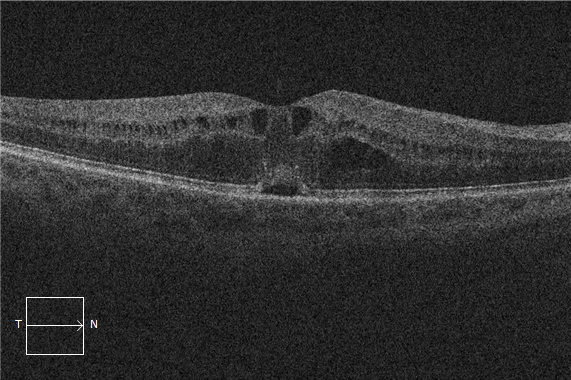
But… in patients with idiopathic macular telangiectasias, it’s not quite as clear.
In these cases, it’s uncertain what’s inside the cyst. Many experts believe this is simply neural degeneration and not true fluid accumulation from vascular extravasation.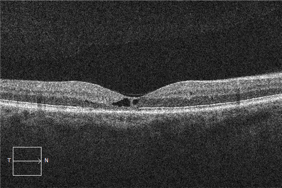
Tip: Call it “intraretinal fluid” and call it a day
As you become more and more of a guru in OCT interpretation, you will use more descriptive terminologies for suggesting diagnoses. In the meantime, if you’re not ready to commit to subtle differentiations, we suggest using the term “intraretinal fluid.” Why? Because the most dangerous and vision-threatening aspect would be intraretinal fluid. So if you call it “intraretinal fluid,” ask for a second opinion and it turns out to be a benign finding, then no harm, no foul. But, if you miss intraretinal fluid by calling it “an inactive neural degeneration,” you may be missing a treatable pathology. Of course, when it comes to your patients and your practice, it’s ultimately up to you. But our position is that it’s better to be safe than sorry. Always learning, The EyeCarePD TeamA Clear Vision for Professional Development
Elevate your eye care skills with image-rich content and relevant case examples that empower you to treat patients more effectively.
Send this Newsletter to a Friend