EyeCarePD

A Clear Vision for Professional Development

  • Catalog
  • FAQ
  • Blog
  • About
  • Contact Us
  • English
    • Português
    • Français
  • Log in

Vitreomacular Adhesion (VMA)

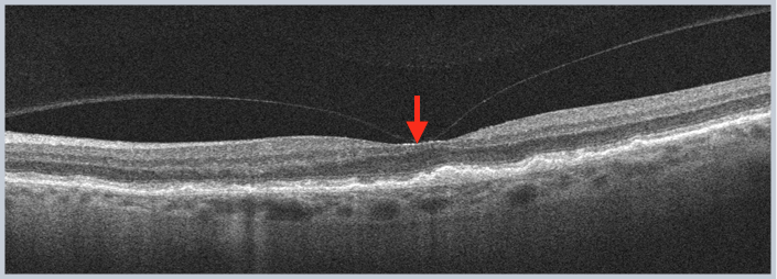
This terms refers to evidence of macular attachment of the posterior cortical vitreous within a central 3-mm radius of the fovea and evidence of perifoveal posterior cortical vitreous detachment. Critically, there is no detectable change in the foveal contour or underlying retinal tissue.

VMA

Practice identifying Vitreomacular Adhesion vs. Vitreomacular Traction now!
« Full-Thickness Macular Hole (FTMH)
Vitreomacular Traction (VMT) »
  • Macular OCT Glossary
    • Retinal Pigment Epithelium (RPE)
    • Ellipsoid Zone (EZ)
    • Subretinal Fluid (SRF)
    • Pigment Epithelial Detachment (PED)
    • Atrophy
    • Subretinal Hyperreflective Material (SHM)
      • Amorphous SHM
      • Consolidated SHM
    • Intraretinal Fluid (IRF)
      • IRF: Empty Cysts
      • IRF: Speckled
      • IRF: Diffuse Outer Thickening
      • IRF: Intraretinal Cystoid Lesions
    • Inner Retinal Opacification
    • Hyperreflective Foci
    • Outer Retinal Tubulations (ORT)
    • Posterior Cortical Vitreous
    • Epiretinal Membrane (ERM)
    • Lamellar Macular Hole (LMH)
    • Full-Thickness Macular Hole (FTMH)
    • Vitreomacular Adhesion (VMA)
    • Vitreomacular Traction (VMT)
    • Macular Schisis
  • About EyeCarePD
  • Blog
  • FAQ
  • Catalog
  • Contact support
  • Macular OCT Glossary

EyeCarePD Inc.
All Rights Reserved

By using this site you agree
to our Terms and Conditions.

Privacy & Cookies Policy

  • Login
  • Sign Up
Forgot Password?
Lost your password? Please enter your username or email address. You will receive a link to create a new password via email.
body::-webkit-scrollbar { width: 7px; } body::-webkit-scrollbar-track { border-radius: 10px; background: #f0f0f0; } body::-webkit-scrollbar-thumb { border-radius: 50px; background: #dfdbdb }