EyeCarePD

A Clear Vision for Professional Development

  • Catalog
  • FAQ
  • Blog
  • About
  • Contact Us
  • English
    • Português
    • Français
  • Log in

Pigment Epithelial Detachment (PED)

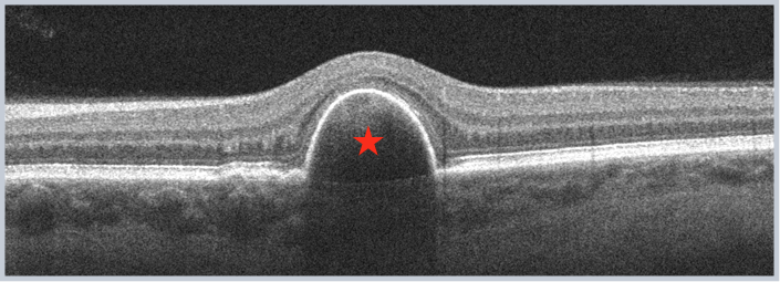
This area is visible as a sharply demarcated dome-shaped retinal pigment epithelial (RPE) elevation. Classically, three main types of PEDs are noted: a) drusenoid, b) serous, and c) vascularized. There is some controversy about these terms and some clinicians prefer to categorize PEDs as: a) vascular or b) avascular.

PED

Practice identifying Pigment Epithelium Detachment now!
« Subretinal Fluid (SRF)
Atrophy »
  • Macular OCT Glossary
    • Retinal Pigment Epithelium (RPE)
    • Ellipsoid Zone (EZ)
    • Subretinal Fluid (SRF)
    • Pigment Epithelial Detachment (PED)
    • Atrophy
    • Subretinal Hyperreflective Material (SHM)
      • Amorphous SHM
      • Consolidated SHM
    • Intraretinal Fluid (IRF)
      • IRF: Empty Cysts
      • IRF: Speckled
      • IRF: Diffuse Outer Thickening
      • IRF: Intraretinal Cystoid Lesions
    • Inner Retinal Opacification
    • Hyperreflective Foci
    • Outer Retinal Tubulations (ORT)
    • Posterior Cortical Vitreous
    • Epiretinal Membrane (ERM)
    • Lamellar Macular Hole (LMH)
    • Full-Thickness Macular Hole (FTMH)
    • Vitreomacular Adhesion (VMA)
    • Vitreomacular Traction (VMT)
    • Macular Schisis
  • About EyeCarePD
  • Blog
  • FAQ
  • Catalog
  • Contact support
  • Macular OCT Glossary

EyeCarePD Inc.
All Rights Reserved

By using this site you agree
to our Terms and Conditions.

Privacy & Cookies Policy

  • Login
  • Sign Up
Forgot Password?
Lost your password? Please enter your username or email address. You will receive a link to create a new password via email.
body::-webkit-scrollbar { width: 7px; } body::-webkit-scrollbar-track { border-radius: 10px; background: #f0f0f0; } body::-webkit-scrollbar-thumb { border-radius: 50px; background: #dfdbdb }