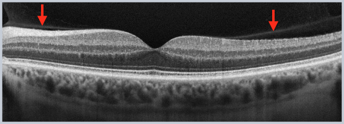
This terms refers to the dense collagen structure that contains the vitreous body. In youth, the posterior cortical vitreous is firmly attached to the internal limiting membrane of the retina and may not be detected on OCT imaging. Additionally, after a complete posterior vitreous detachment has occurred, the posterior cortical vitreous may shift upward on the scan and may not be visible. Only during periods of partial release from the retina will the posterior cortical vitreous be reliably detected on OCT.
