EyeCarePD

A Clear Vision for Professional Development

  • Catalog
  • FAQ
  • Blog
  • About
  • Contact Us
  • English
    • Português
    • Français
  • Log in

IRF: Diffuse Outer Thickening

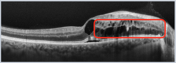
This subtype of IRF is characterized by hyporeflective ovoid spaces that are principally located in the outer half of the retina.

IRF Diffuse Outer Thickening

Practice identifying Intraretinal Fluid: Diffuse Outer Thickening now!
« IRF: Speckled
IRF: Intraretinal Cystoid Lesions »
  • Macular OCT Glossary
    • Retinal Pigment Epithelium (RPE)
    • Ellipsoid Zone (EZ)
    • Subretinal Fluid (SRF)
    • Pigment Epithelial Detachment (PED)
    • Atrophy
    • Subretinal Hyperreflective Material (SHM)
      • Amorphous SHM
      • Consolidated SHM
    • Intraretinal Fluid (IRF)
      • IRF: Empty Cysts
      • IRF: Speckled
      • IRF: Diffuse Outer Thickening
      • IRF: Intraretinal Cystoid Lesions
    • Inner Retinal Opacification
    • Hyperreflective Foci
    • Outer Retinal Tubulations (ORT)
    • Posterior Cortical Vitreous
    • Epiretinal Membrane (ERM)
    • Lamellar Macular Hole (LMH)
    • Full-Thickness Macular Hole (FTMH)
    • Vitreomacular Adhesion (VMA)
    • Vitreomacular Traction (VMT)
    • Macular Schisis
  • About EyeCarePD
  • Blog
  • FAQ
  • Catalog
  • Contact support
  • Macular OCT Glossary

EyeCarePD Inc.
All Rights Reserved

By using this site you agree
to our Terms and Conditions.

Privacy & Cookies Policy

  • Login
  • Sign Up
Forgot Password?
Lost your password? Please enter your username or email address. You will receive a link to create a new password via email.
body::-webkit-scrollbar { width: 7px; } body::-webkit-scrollbar-track { border-radius: 10px; background: #f0f0f0; } body::-webkit-scrollbar-thumb { border-radius: 50px; background: #dfdbdb }