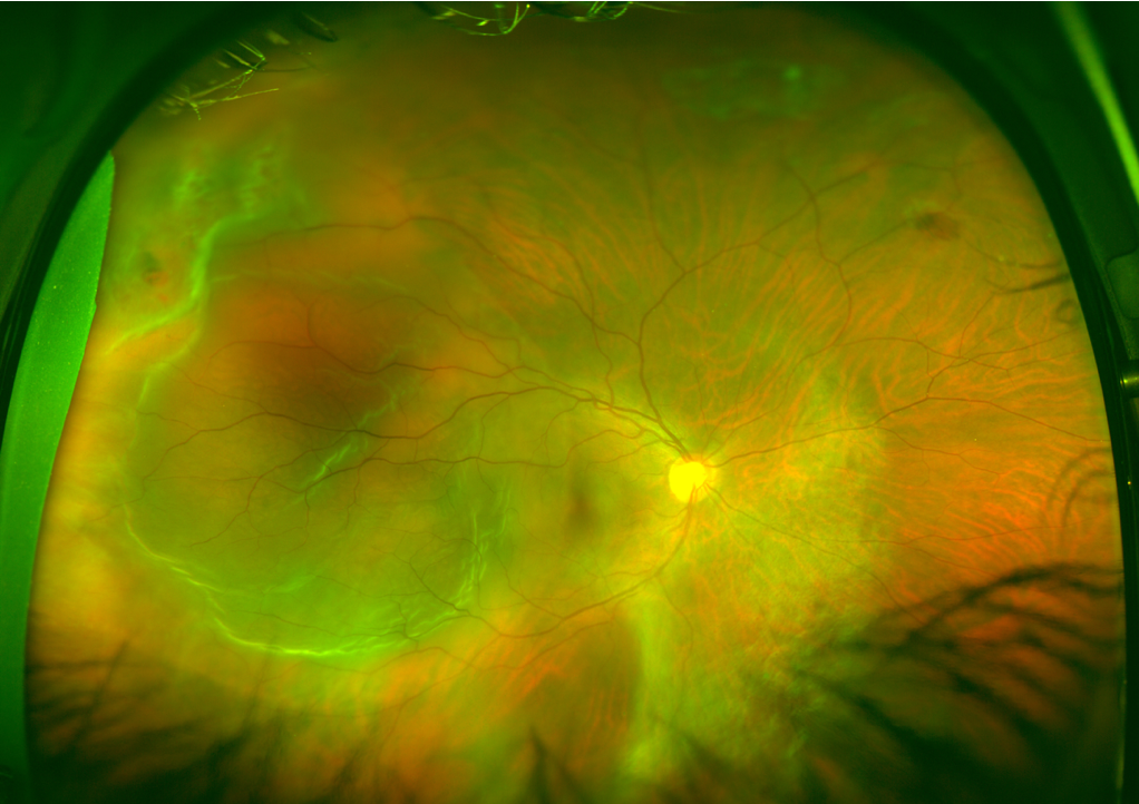
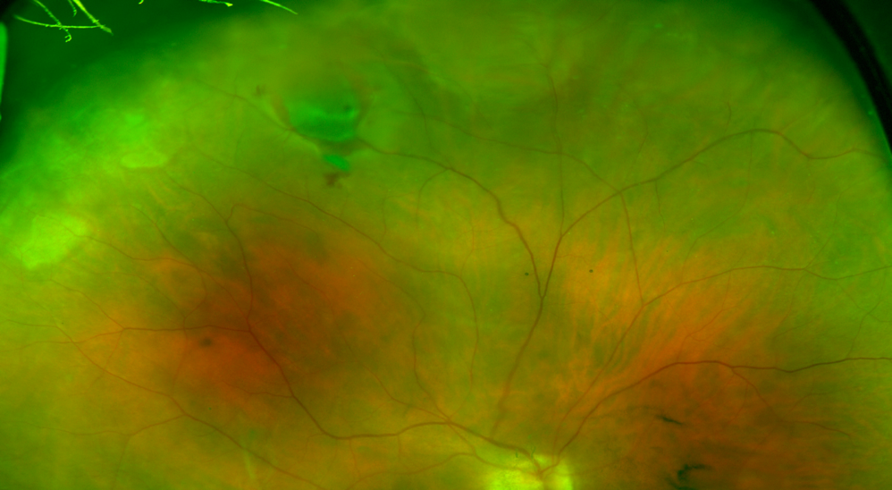
43-year-old woman with 2 adjacent retinal breaks (red circle) and prominent subretinal fluid that involves the macula (blue circle).
A Clear Vision for Professional Development
43-year-old woman with 2 adjacent retinal breaks (red circle) and prominent subretinal fluid that involves the macula (blue circle).

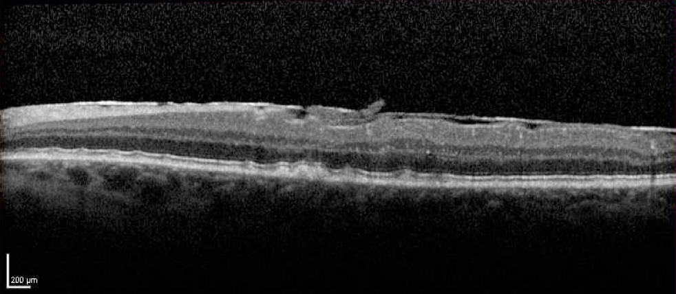
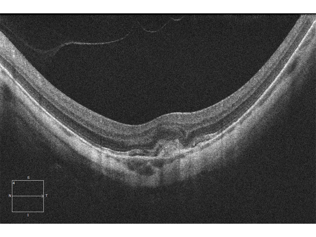
Are you able to find the SHM and SRF?
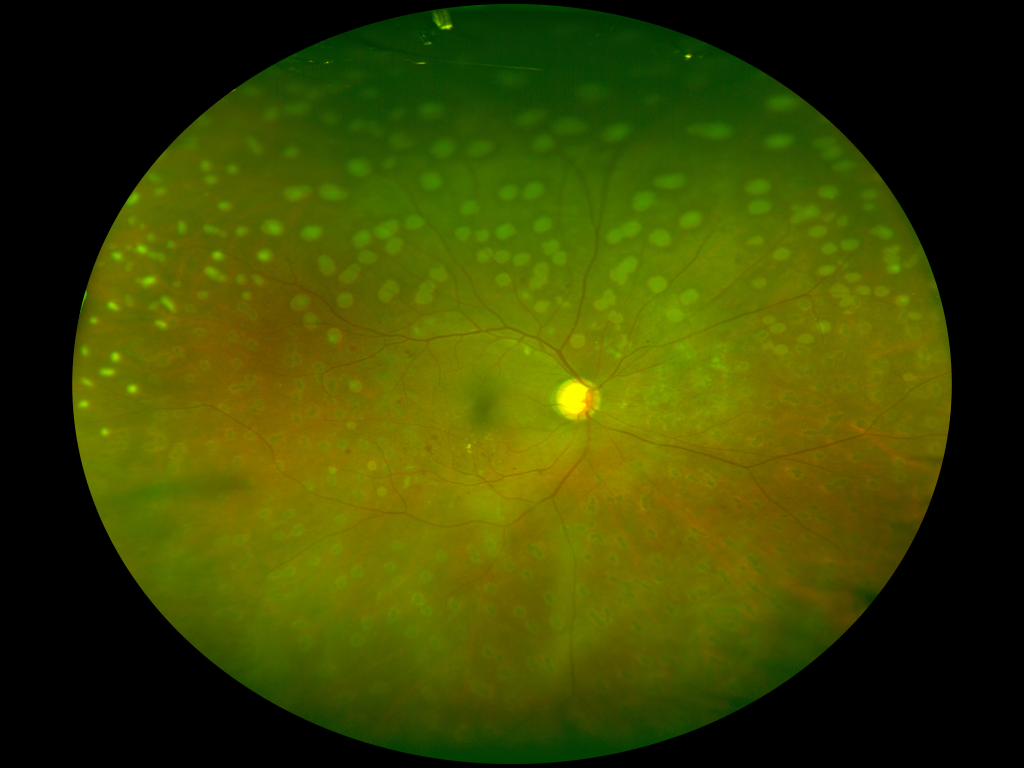
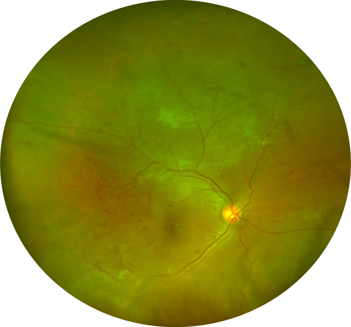
61-year-old man with known dry AMD on routine follow-up examination.
61-year-old man, status post PRP. The inferior laser demonstrates 4-week-old laser burns.

63-year-old man with clinically visible asteroid hyalosis.
60-year-old man with an embolic BRAO. There was typical retinal whitening on clinical examination. The OCT demonstrates the area of the occlusion with an abrupt transition from the normal to abnormal retina. While not always required, the OCT can prove to be nearly pathognomonic in challenging cases.
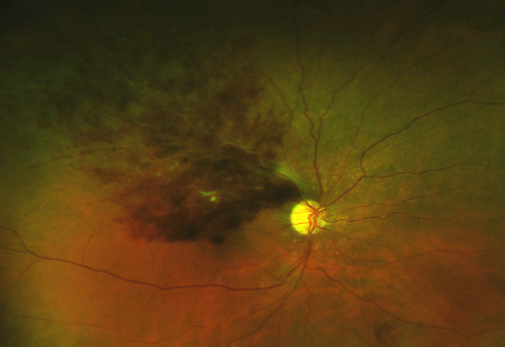
56-year-old man with a horseshoe retinal tear following a spontaneous posterior vitreous detachment.
39-year-old man with multiple large areas of neovascularization secondary to diabetic retinopathy.
58-year-old woman with obvious macular blood and retinal thickening.
42-year-old woman with choroidal neovascularization secondary to myopia.