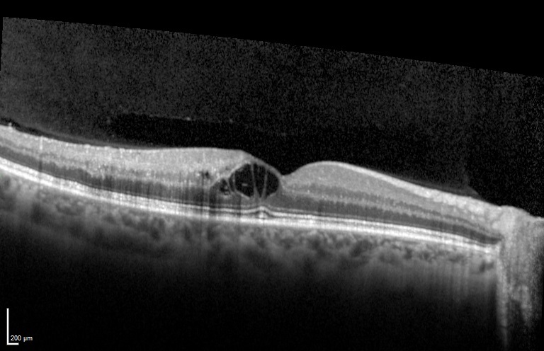
This OCT image is typical of cystoid macular edema after a retinal vein occlusion.
A Clear Vision for Professional Development
This OCT image is typical of cystoid macular edema after a retinal vein occlusion.
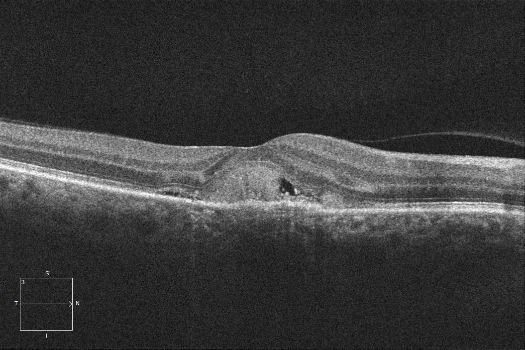
This OCT image is typical of choroidal neovascularization.
43-year-old man with no known past medical history presents for a routine eye exam.
83-year-old man with Dry Age Related Macular Degeneration and an Epiretinal Membrane.
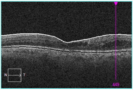
73-year-old man with diabetic retinopathy and prior panretinal photocoagulation for proliferative disease that is now regressed and stable.
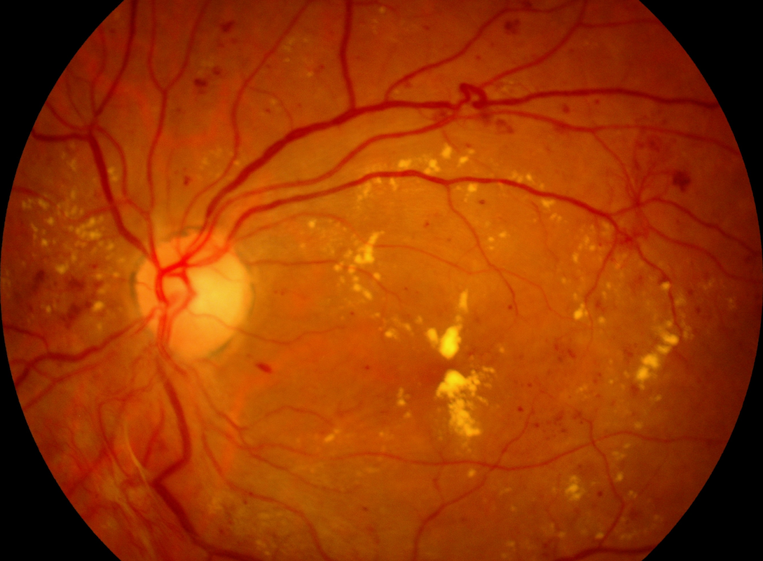
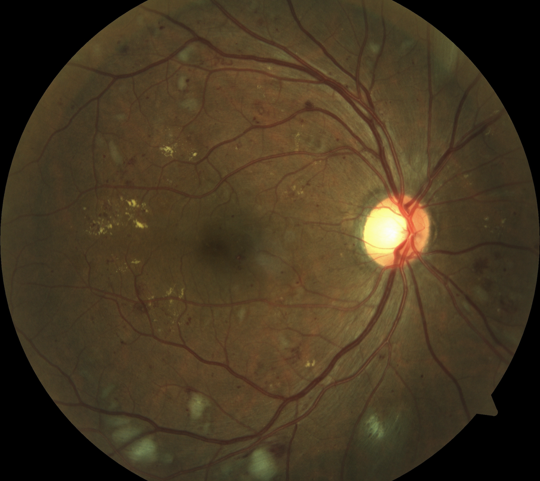
This color fundus photograph demonstrates proliferative diabetic retinopathy with clinically significant macular edema.
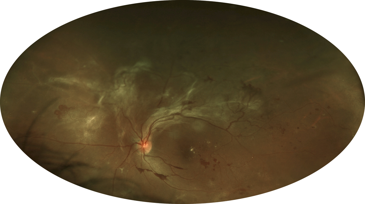
54-year-old woman who presented initially with active proliferative diabetic retinopathy and fibrotic changes.
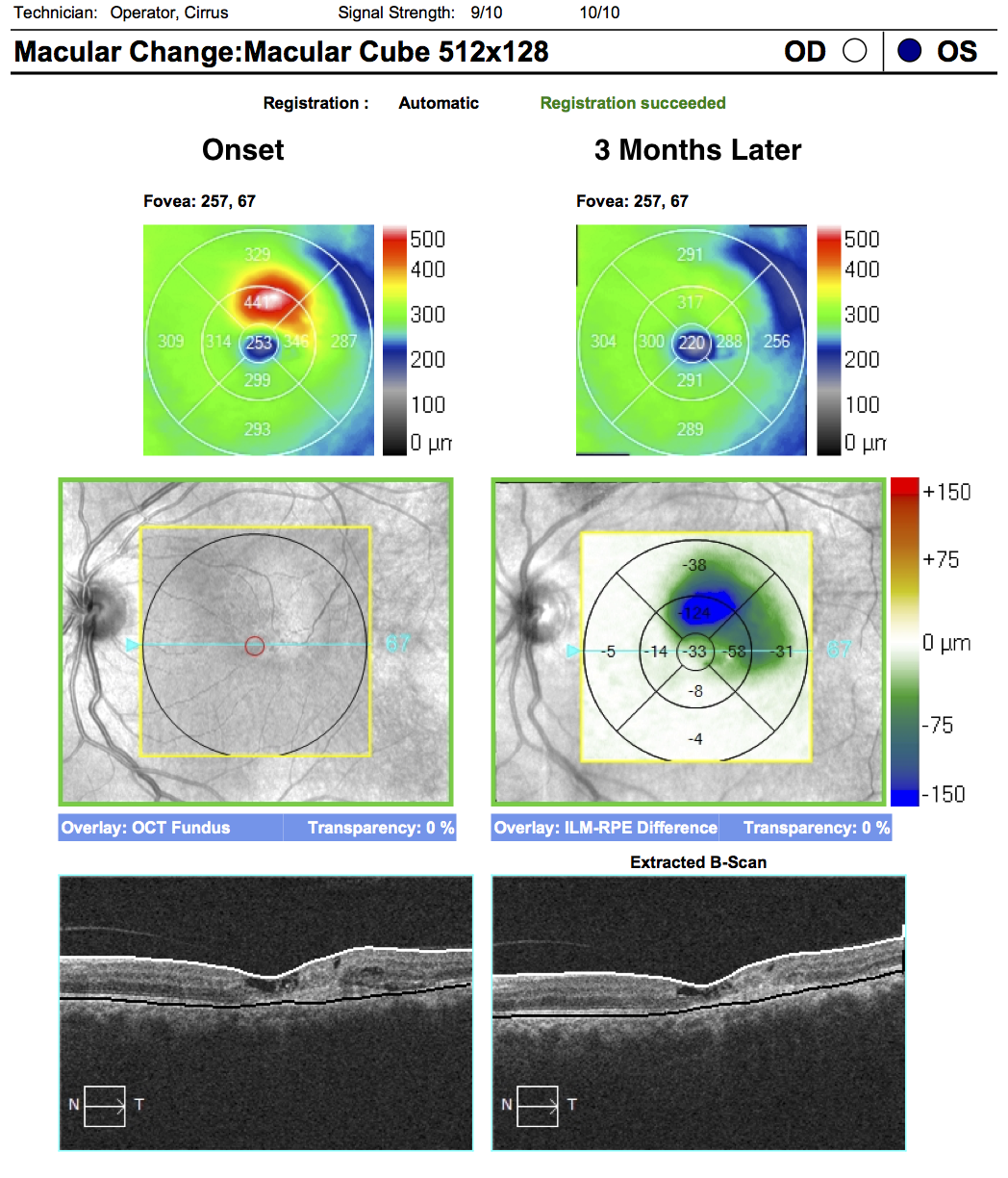
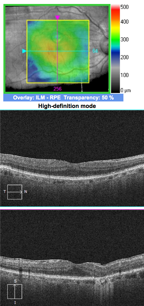
62-year-old woman with 3 monthly anti-VEGF injections. The images presented compare the presenting OCT to the follow-up OCT.
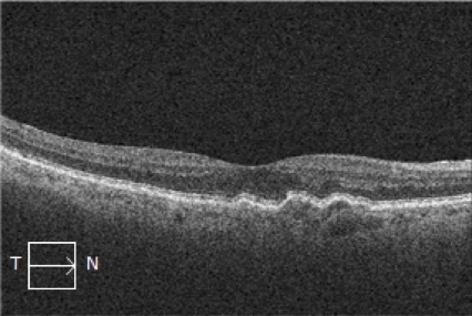
69-year-old man with Wet Age Related Macular Degeneration who is stable on quarterly anti-VEGF intravitreal injections presents for a routine follow-up evaluation.
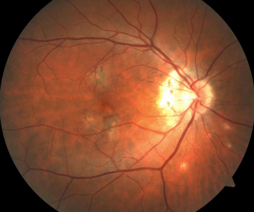
36-year-old man with choroidal neovascularization (CNV) secondary to presumed ocular histoplasmosis.