Vitreous degeneration can take varying forms and has been classified in various manners in the scientific literature.
A Clear Vision for Professional Development
Vitreous degeneration can take varying forms and has been classified in various manners in the scientific literature.

We certainly all would agree that not every area of hard exudate warrants intervention.
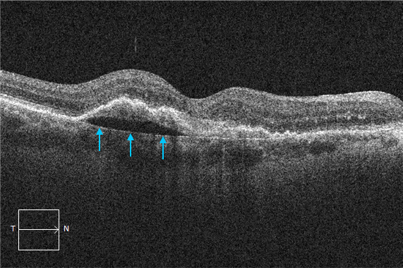
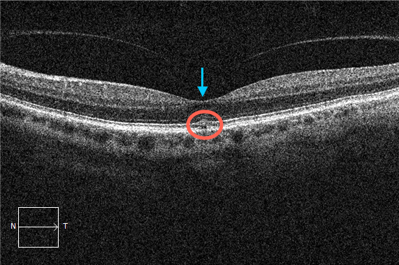
In this OCT image you will note an epiretinal membrane (arrows). There is also intraretinal fluid best described as diffuse outer retinal thickening (circle).
OCT interpretation relies on correctly identifying several key structures. One of these is the retinal pigment epithelium (RPE).
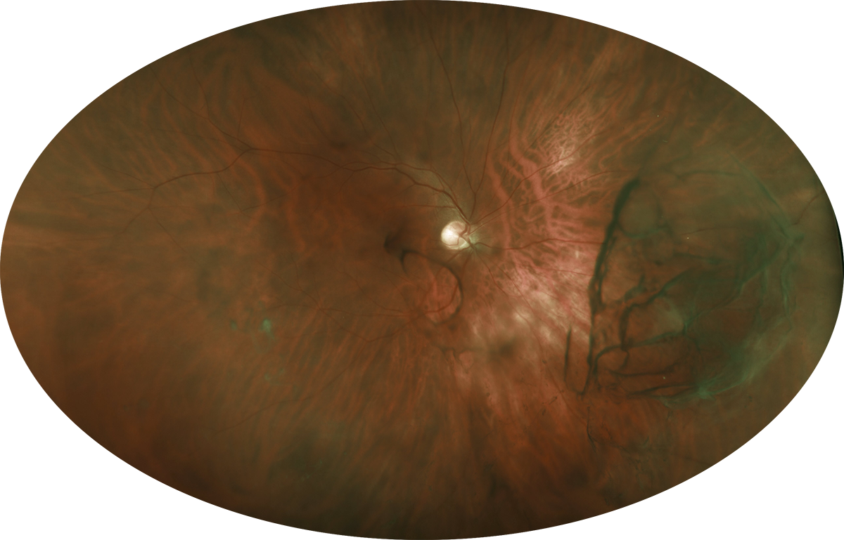
This was an incidental finding in a 60-year-old African-American female.
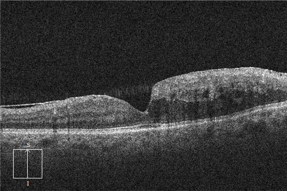
Full thickness macular hole formation commences in the outer retina and progresses inward.
OCT evaluation is a skill in pattern recognition. While this is not an unusual concept for eye care professionals this is not commonly discussed in terms of OCT interpretation.
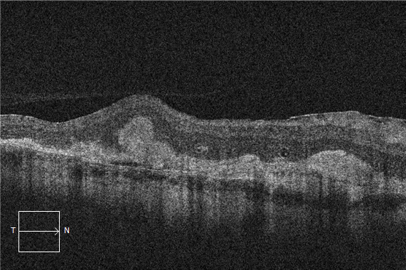
ORT stands for outer retinal tubulation. It is theorized to be due to degenerated photoreceptors that become arranged in a circular or ovoid pattern.
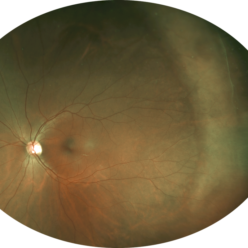
The image displayed is not a diffuse amelanotic choroidal melanoma.
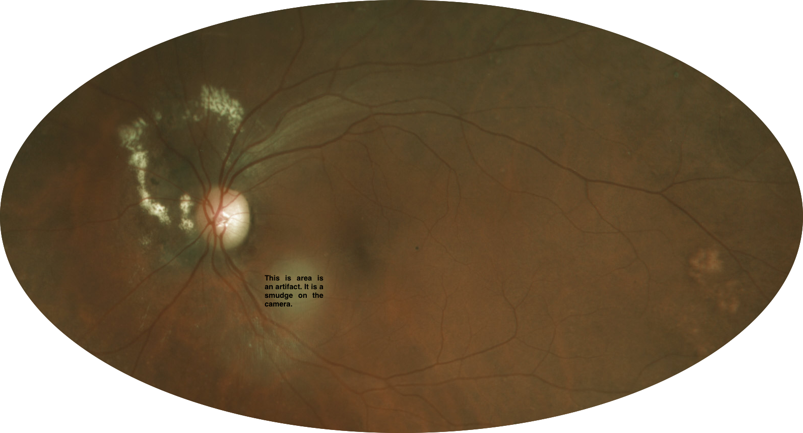
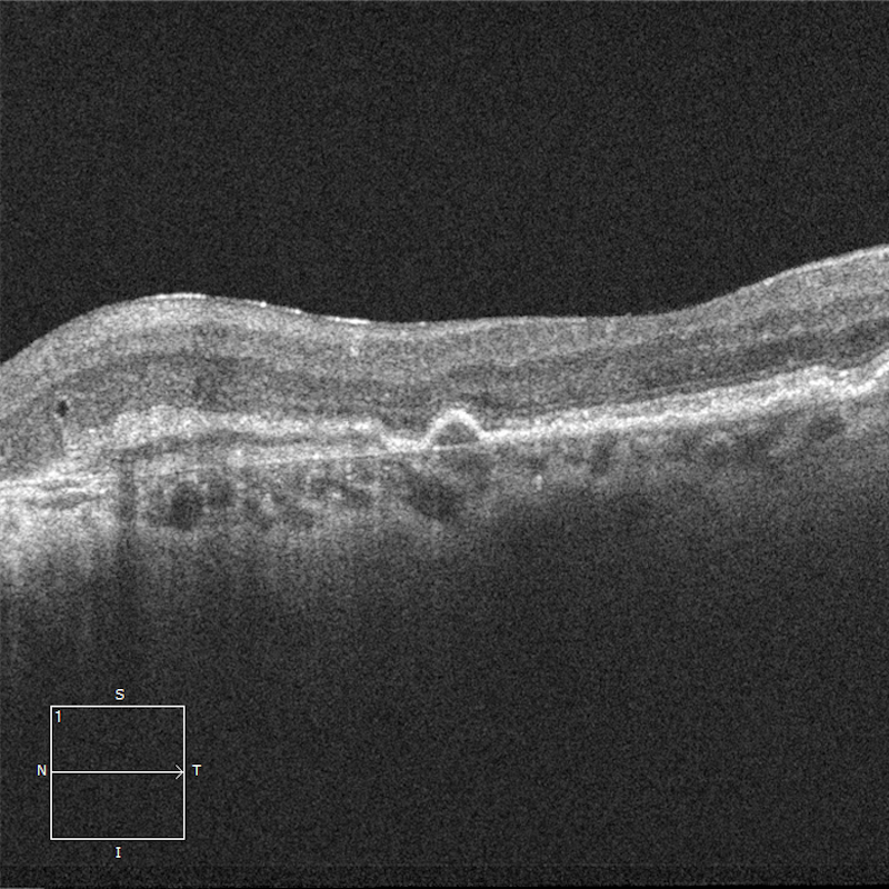
This OCT image is typical of dry age-related macular degeneration complicated by focal atrophy.