EyeCarePD

A Clear Vision for Professional Development

  • Catalog
  • Blog
  • About
  • Contact Us
  • English
  • Log in

When is it bad to be “bright”?

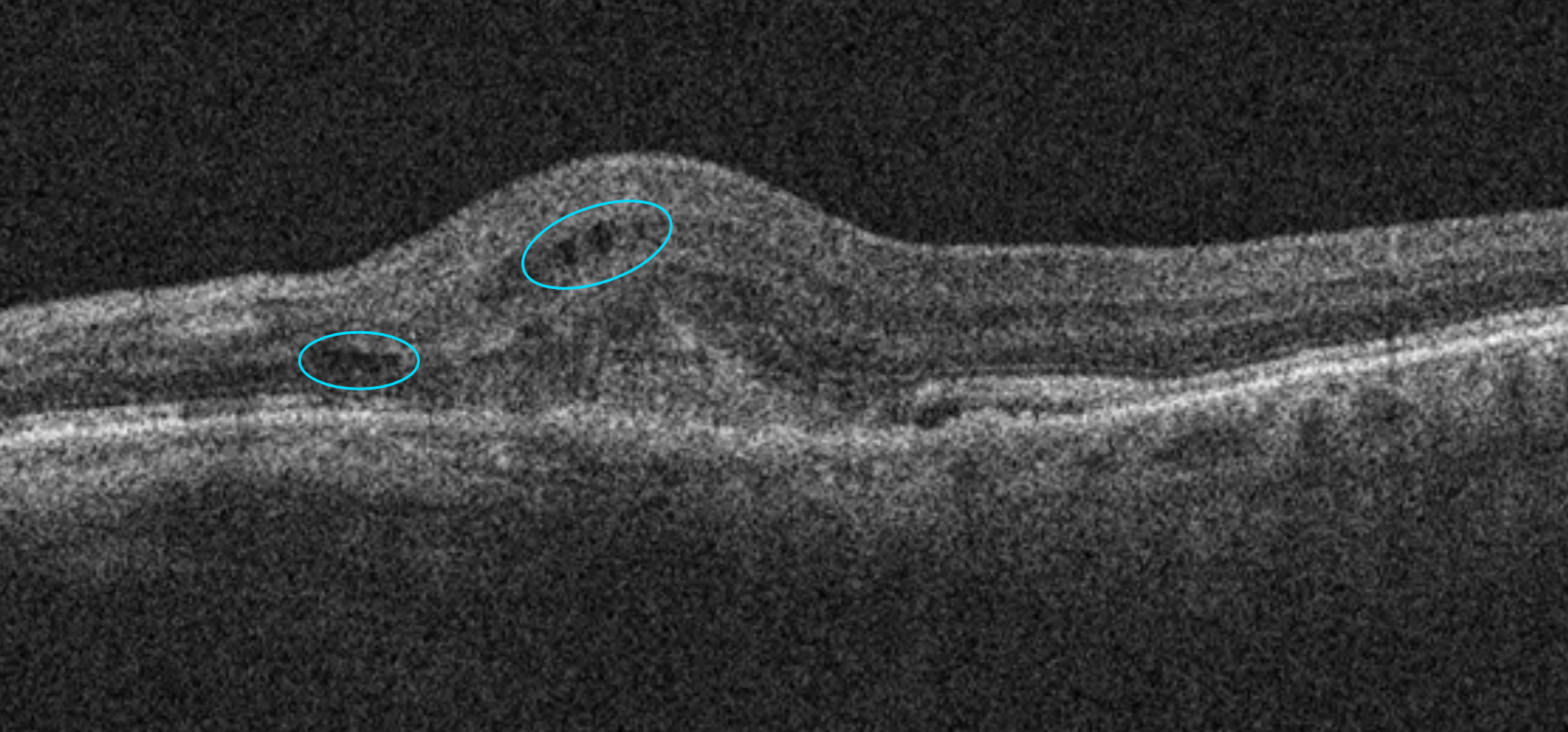
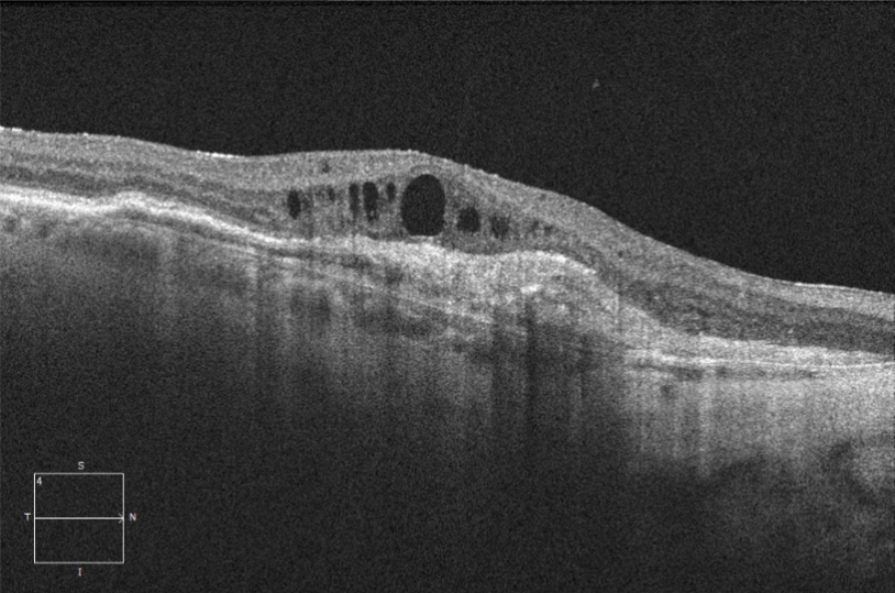
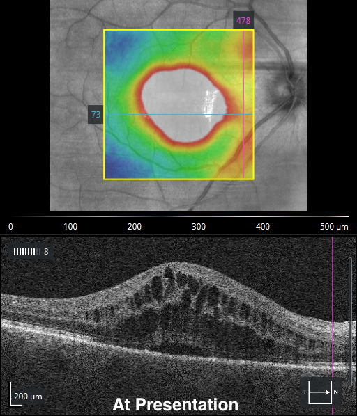
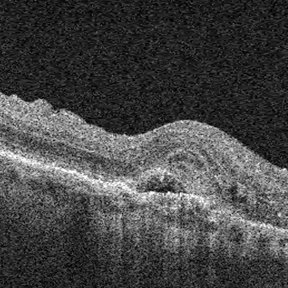
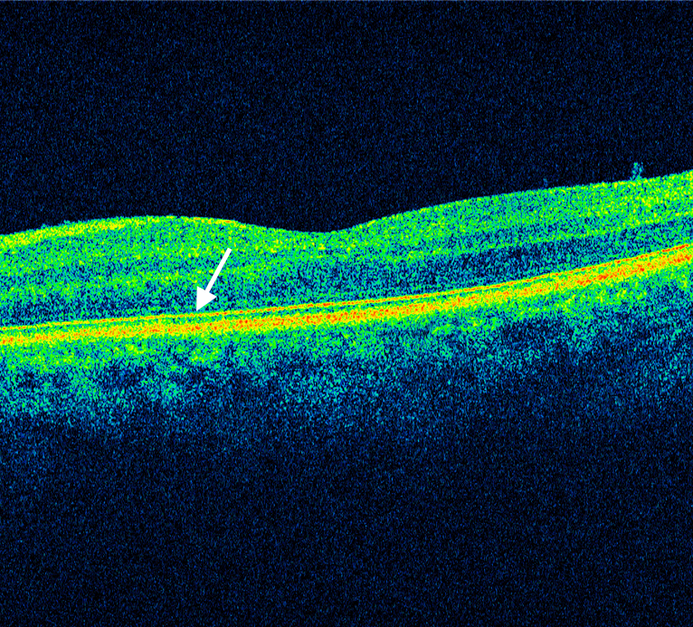
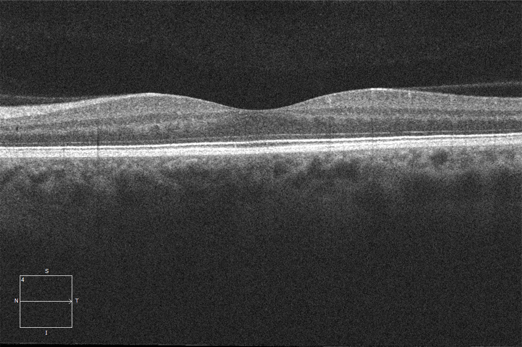
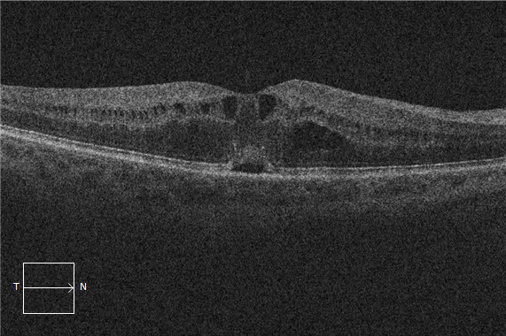
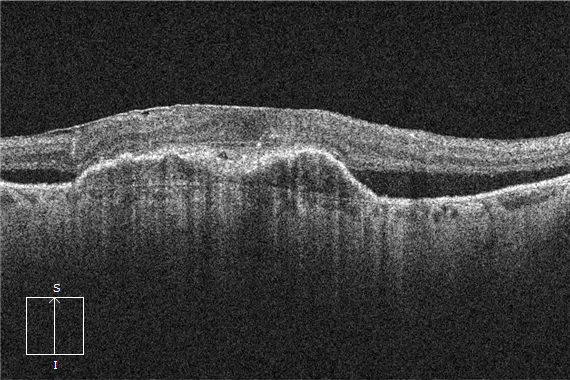
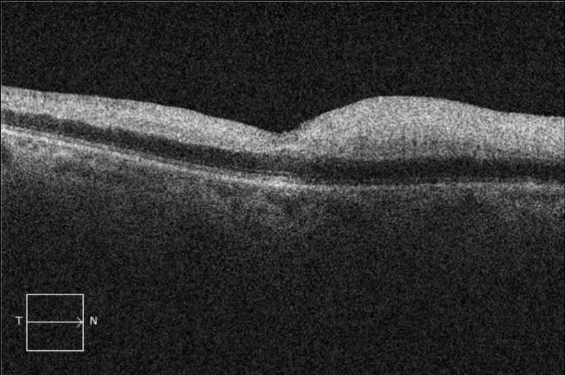
This edition of the EyeCarePD newsletter is about optical intensity and Retinal Artery Occlusions. Let’s talk about Retinal Arterial Occlusion (RAO), a vision-threatening pathology with visual outcomes ranging from 20/25 to no light perception. Even though we can often diagnosis RAO on clinical examination, adjunctive OCT imaging can still be useful. That’s because the OCT… Read More

Is it really RAP? And why you should care.

This edition of the EyeCarePD newsletter is about retinal angiomatous proliferation and how it affects patient care. Want to learn about an interesting variant of Wet Age-Related Macular Degeneration (AMD)? Let’s talk about retinal angiomatous proliferation (RAP). Unlike choroidal neovascularization (CNV), which starts in the choroid, RAP starts in the retina. The neovessels then proliferate… Read More

Does your patient need to be seen by a specialist ASAP?

This edition of the EyeCarePD newsletter is about timeframes for patient referrals for common macular pathologies. We’ve all seen patients with concerning presentations of macular disease. From blood to fluid, it can be confusing for the referring doctor to know how urgently a patient needs to be seen by a specialist. At EyeCarePD, we’re often… Read More

A new term is born intraretinal cystoid lesions

There just isn’t enough information out there when it comes to OCT terminology for intraretinal fluid—specifically, for the purpose of lesion classification in chorioretinal pathologies.

Treating postoperative cystoid macular edema: then and now

This edition of the EyeCarePD newsletter introduces new technology for improving the accuracy of OCT interpretation and highlights advances in the treatment of postoperative cystoid macular edema.

What a difference a little contrast can make

OCT Optical Coherence Tomography

This edition of the EyeCarePD newsletter is about the importance of viewing macular OCT images with different contrast and background color settings.

Pseudocolor or grayscale? That is the question.

This edition of the EyeCarePD newsletter is about the thousands of levels of intensity detected by an OCT device, as well as the difference between viewing images in pseudocolor and grayscale.

What you need to know about inner retinal opacification

Inner retinal opacification is a common finding seen on OCTs for retinal artery occlusions.

Again with the fluid?

Let’s continue the conversation about fluid. But this time, the spotlight is on intraretinal fluid.

Wait, is that really subretinal fluid?

In this newsletter, Dr. David Lederer, CEO of EyeCarePD explains why you should use the term “subretinal fluid” first, before committing to subtle differentiations.

« Previous Page
Next Page »

For past issues, checkout the newsletter archive.

  • About EyeCarePD
  • Blog
  • Contact support
  • Macular OCT Glossary

EyeCarePD Inc.
All Rights Reserved

By using this site you agree
to our Terms and Conditions.

Privacy & Cookies Policy