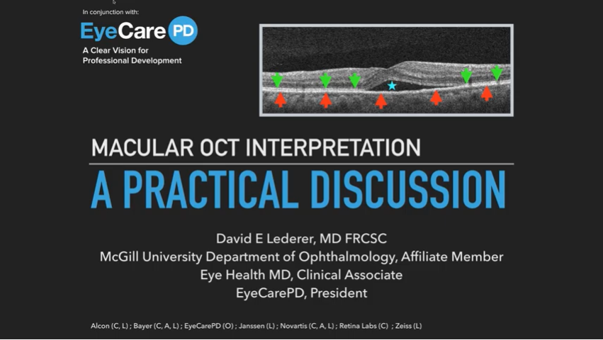
The EyeCarePD educational platform is designed to promote active learning through interaction with OCTs. That said, I still enjoy delivering traditional lectures from time to time. I’d like to share some of these with you. Here is one I gave at a trade show a while back. The original post had 145k views on the… Read More
Retina Hot Mic Case Series 02
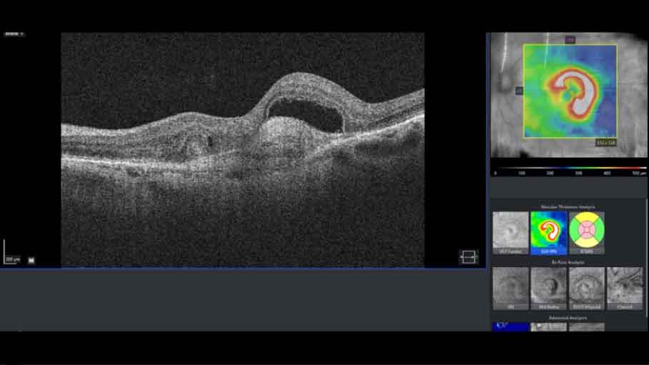
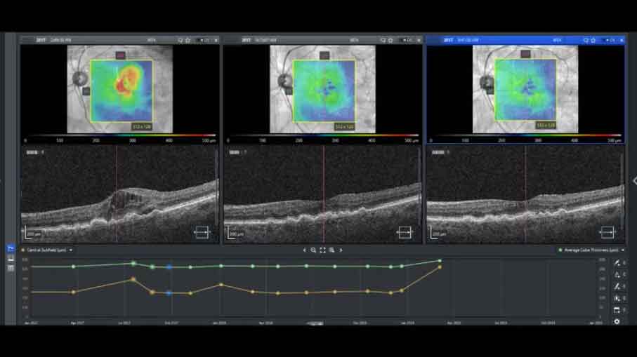
ERM and CNV. Can you see it? Watch Now…
Retina Hot Mic Case Series 01
Welcome to our new Retina Hot Mic series, where you get to look over Dr. David’s shoulder as he interprets retinal imagery and explains his thinking process as he refines his diagnosis. Watch Now…
Green Is Not Always Good
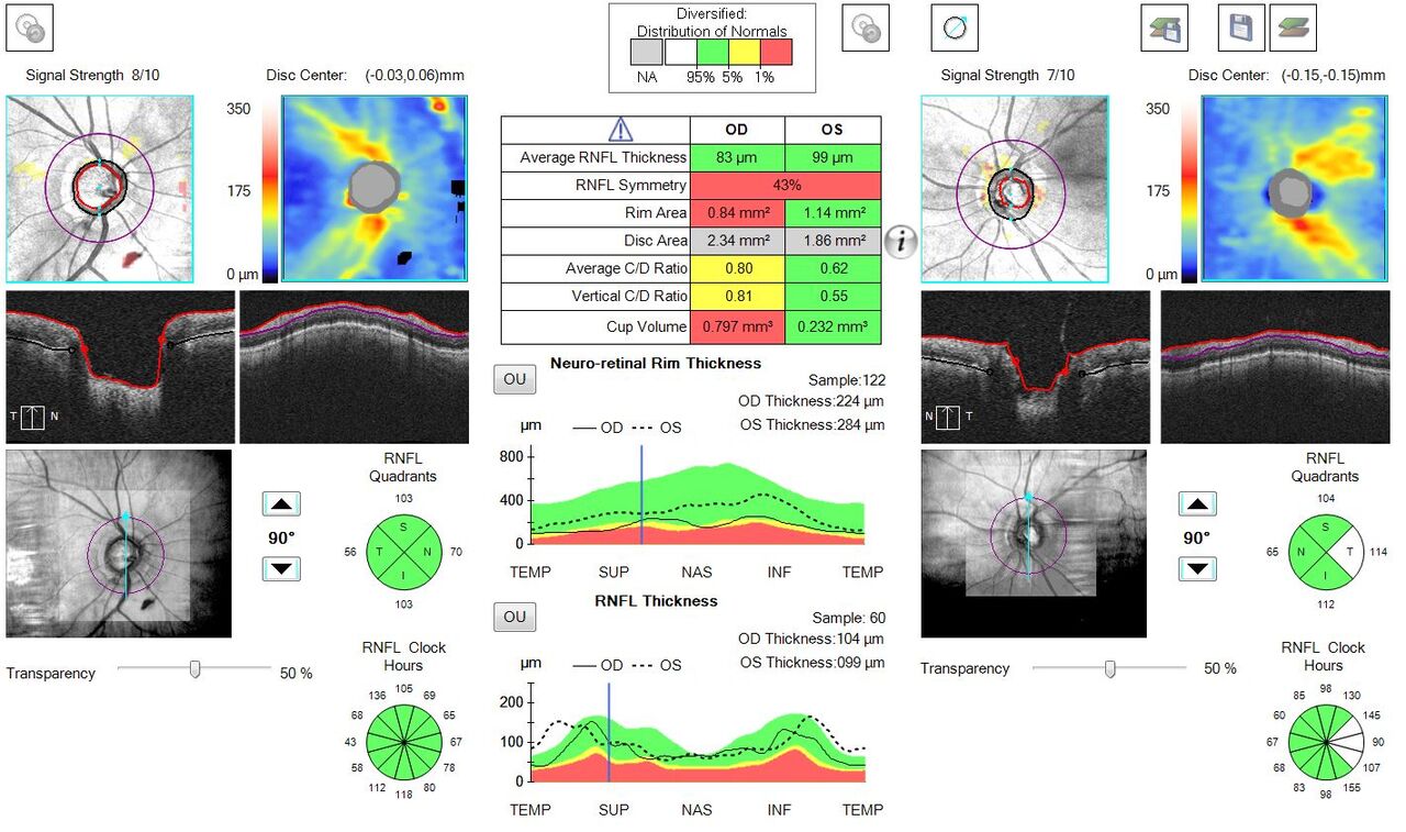
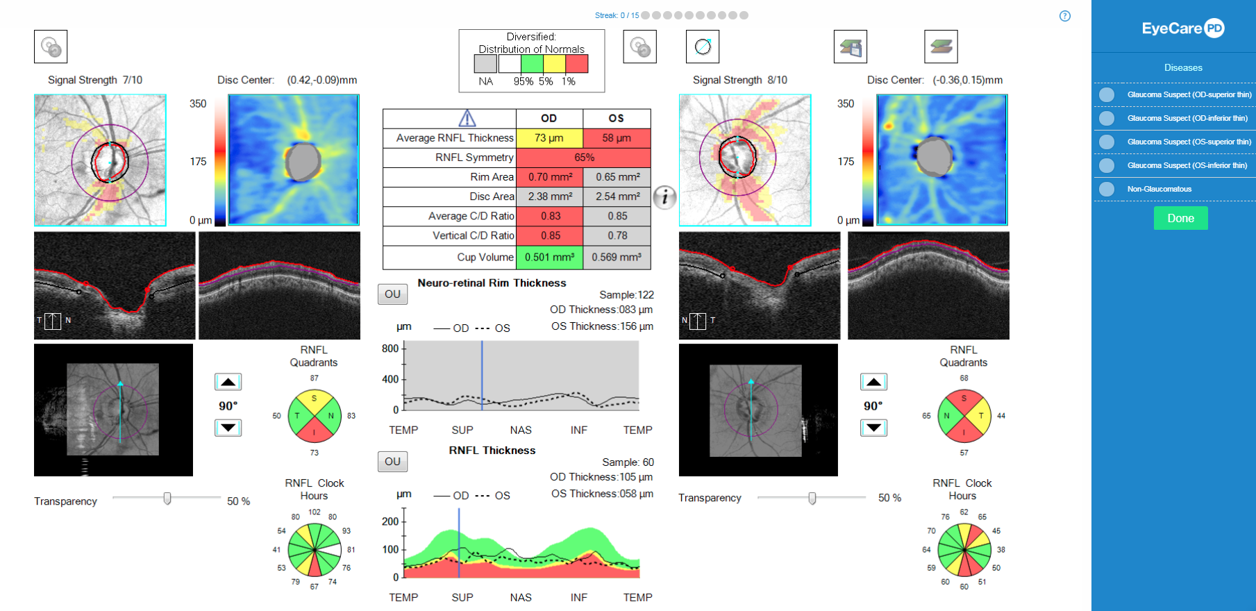
This edition of the EyeCarePD newsletter is about understanding the strengths and limitations of the RNFL analysis when considering a diagnosis of glaucoma. The INTERPRET: Glaucoma training software, which many of you have had a chance to play, is all about learning and practicing common skills needed to diagnose disease. However, we also all know… Read More
The Devil Is In The Details
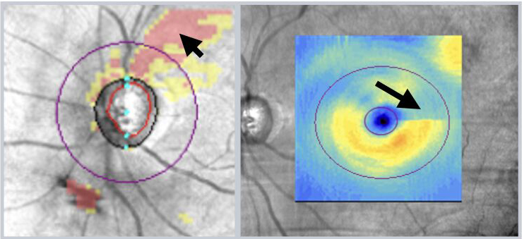
This edition of the EyeCarePD Newsletter digs deeper into issues to think about when considering the utility of OCT to evaluate your glaucoma patients. With our recent post discussing the 2017 publication from the Advanced Imaging For Glaucoma Study Group, we thought now would be a good time to pick out a few of the… Read More
The OCT-VF-Glaucoma Relationship
This edition of the EyeCarePD Newsletter provides the basic background you need to successfully use OCT to evaluate your glaucoma patients. How do you follow your patients who are Glaucoma Suspects? What about those with definitive Glaucoma? Does your algorithm vary by the extent of disease (mild, moderate, severe)? In a seminal publication by Zhang… Read More
Interpret Glaucoma OCT Scans Like a Pro
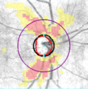
EyeCarePD is proud to announce our latest learning program, INTERPRET: Glaucoma. Using game-based techniques we make learning fun again! Try it now. For free. You need to be good at OCT interpretation to be good at your job. This course will begin your transformation into an OCT expert. After showing you how to find pathologies… Read More
Macular Edema Case Examples
This edition of the EyeCarePD newsletter is about the treatment of clinically significant diabetic macular edema and provides case examples. As discussed in a previous newsletter, all clinically significant macular edema (CSME) is Diabetic Macular Edema (DME), but not all DME is CSME. Furthermore, CSME is commonly divided into center-involving clinically significant macular edema (ci-CSME)… Read More
Diabetic Macular Edema Laser or Injections?
This edition of the EyeCarePD newsletter is about diabetic macular edema and provides insight into critical definitions. Diabetic Macular Edema (DME) is a complex disease process with variable treatment options. Discussions about treatment options could go on and on. But before we get ahead of ourselves, a simple introduction seems prudent. In 1985, with the… Read More
Do you know about the latest vitreo-mathematical trick?
This edition of the EyeCarePD newsletter is about vitreomacular adhesion and expanding how we think about the area of attachment. It’s no secret that the vitreous shifts based on its water:collagen ratio throughout life. When we’re young, the vitreous is firmly attached to the retina via adhesion molecules along the vitreoretinal interface. As we age,… Read More
When is it bad to be “bright”?
This edition of the EyeCarePD newsletter is about optical intensity and Retinal Artery Occlusions. Let’s talk about Retinal Arterial Occlusion (RAO), a vision-threatening pathology with visual outcomes ranging from 20/25 to no light perception. Even though we can often diagnosis RAO on clinical examination, adjunctive OCT imaging can still be useful. That’s because the OCT… Read More
Is it really RAP? And why you should care.
This edition of the EyeCarePD newsletter is about retinal angiomatous proliferation and how it affects patient care. Want to learn about an interesting variant of Wet Age-Related Macular Degeneration (AMD)? Let’s talk about retinal angiomatous proliferation (RAP). Unlike choroidal neovascularization (CNV), which starts in the choroid, RAP starts in the retina. The neovessels then proliferate… Read More