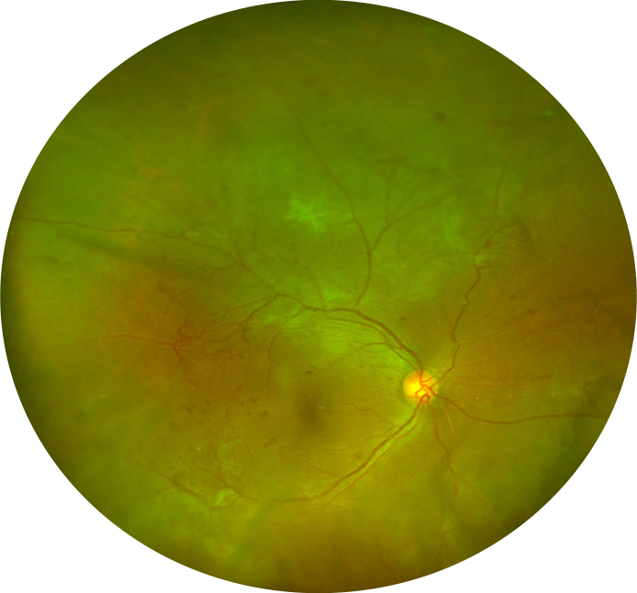
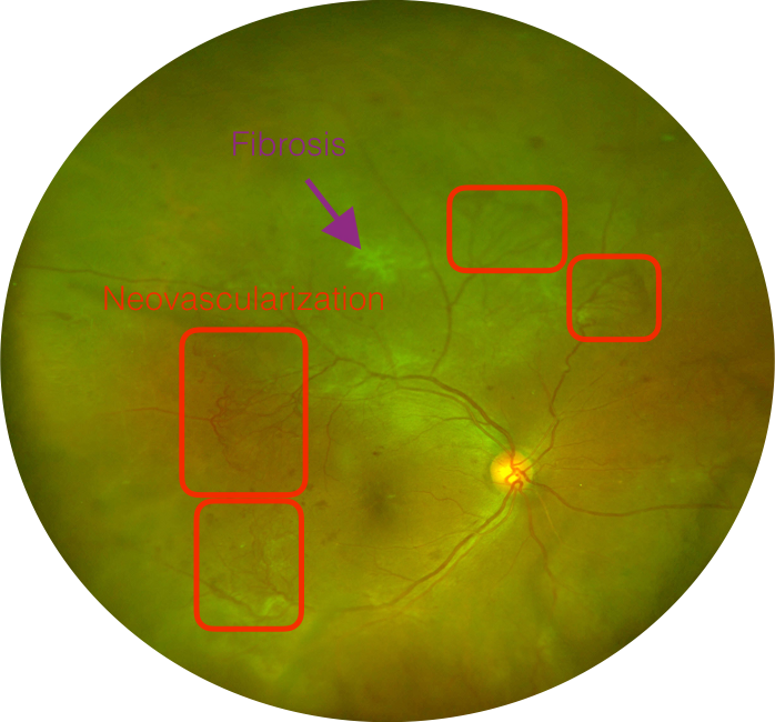
39-year-old man with multiple large areas of neovascularization secondary to diabetic retinopathy. Many of these areas have a fibrotic component (only one area is labelled). This image was obtained with the Optos, and the color variation from what is “normally” seen in a standard flash camera is important to recognize to prevent accidently missing pathology. Nevertheless, this view of the retina is becoming more common as more cSLO devices are employed in routine clinical practice.