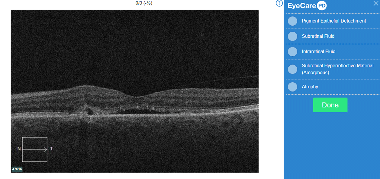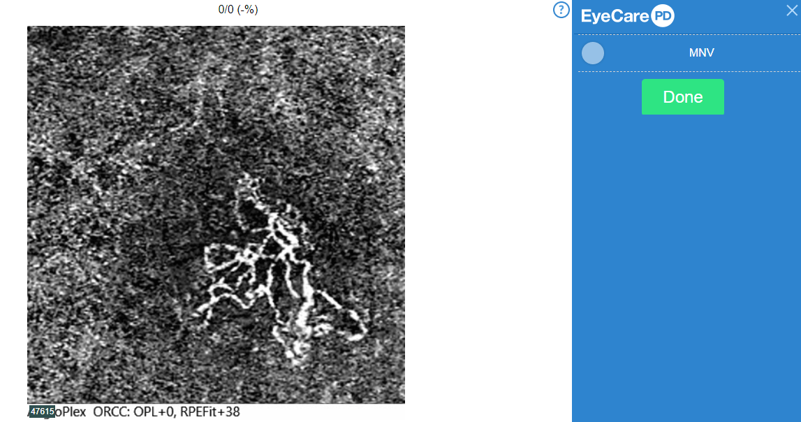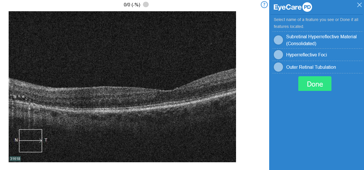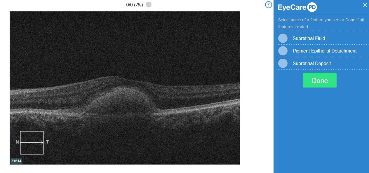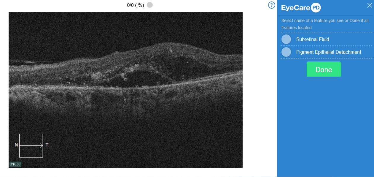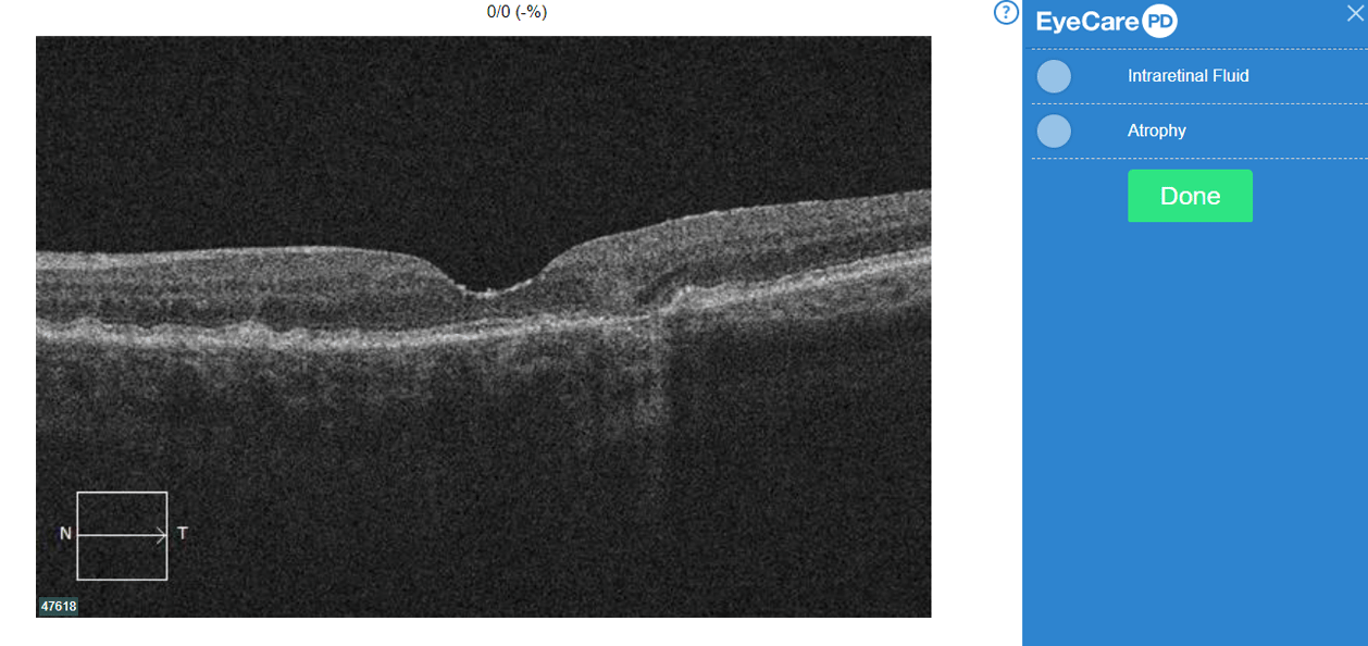
There just isn’t enough information out there when it comes to OCT terminology for intraretinal fluid—specifically, for the purpose of lesion classification in chorioretinal pathologies.
A Clear Vision for Professional Development
There just isn’t enough information out there when it comes to OCT terminology for intraretinal fluid—specifically, for the purpose of lesion classification in chorioretinal pathologies.
This edition of the EyeCarePD newsletter introduces new technology for improving the accuracy of OCT interpretation and highlights advances in the treatment of postoperative cystoid macular edema.
Vitreous degeneration can take varying forms and has been classified in various manners in the scientific literature.
We certainly all would agree that not every area of hard exudate warrants intervention.
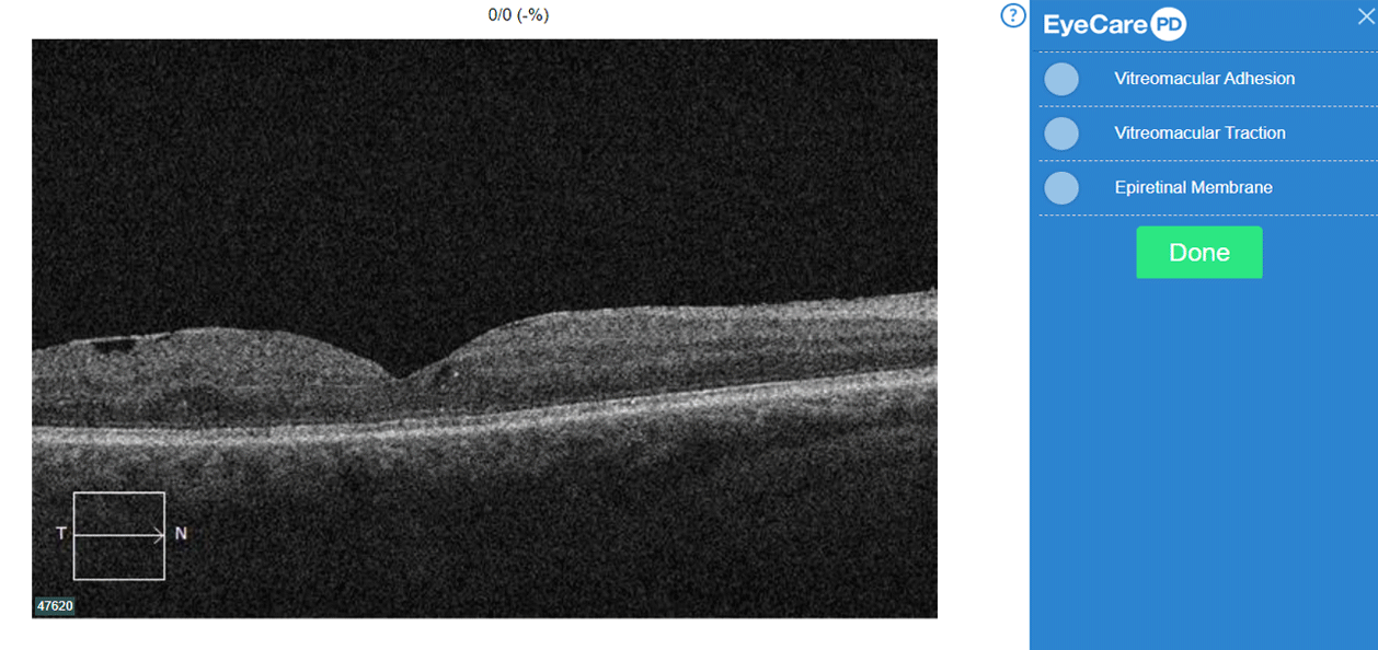
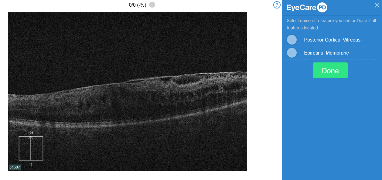
In this OCT image you will note an epiretinal membrane (arrows). There is also intraretinal fluid best described as diffuse outer retinal thickening (circle).
OCT interpretation relies on correctly identifying several key structures. One of these is the retinal pigment epithelium (RPE).
This was an incidental finding in a 60-year-old African-American female.
Full thickness macular hole formation commences in the outer retina and progresses inward.
This edition of the EyeCarePD newsletter is about the importance of viewing macular OCT images with different contrast and background color settings.
OCT evaluation is a skill in pattern recognition. While this is not an unusual concept for eye care professionals this is not commonly discussed in terms of OCT interpretation.
Contact support via email:
support@eyecarepd.com or
telephone 1 (888) 902-2017
EyeCarePD Inc.
All Rights Reserved
By using this site you agree
to our Terms and Conditions.