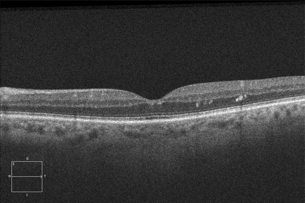
75-year-old man with large central drusen noted clinically.
A Clear Vision for Professional Development
75-year-old man with large central drusen noted clinically.
Diabetic Macular Edema. 75-year-old man with chronic center-involving diabetic macular edema (DME) and a history of anti-VEGF and intravitreal triamcinolone injections. The patient developed a prominent posterior subcapsular cataract (PSCC) that limited the view to the macula. Despite the low-quality OCT signal, the DME is clearly present along with a component of subretinal fluid. Cataract… Read More
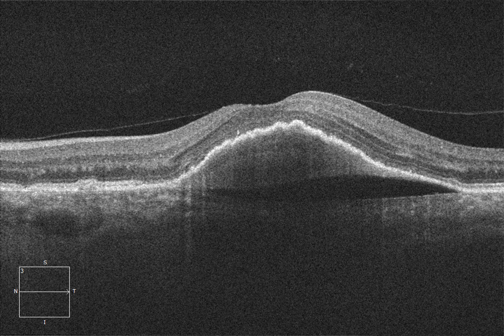
Serous Pigment Epithelial Detachment. 85-year-old female with a large pigment epithelial detachment. Fluorescein angiography did not reveal a choroidal neovascular membrane, and the patient was followed without treatment. After 1 year of follow-up the patient has remained stable without progression.

Wet Age-Related Macular Degeneration. 65-year-old man with choroidal neovascularization as evidenced by subretinal hyperreflective material and subretinal fluid. The back portion of the vitreous (hyaloid) can be seen partially detached. This is a normal finding and should not be confused with vitreomacular traction. Only one treatable pathology is present: wAMD.
Cystoid Fluid. Both patients have cystoid fluid in the macula. Both patients are receiving anti-VEGF injections every 2 months. The OCT can be used to distinguish these pathologies as the patient with wAMD has deep drusen that are absent in the patient with a CRVO. Assessing all the factors on an OCT can be helpful… Read More
Nonproliferative Diabetic Retinopathy without Diabetic Macular Edema. 63-year-old man with mild NPDR. The OCT demonstrates hard exudates, but there is no evidence of macular edema. While hard exudates are a common finding in cases of macular edema, they can occur in isolation and do not warrant treatment.
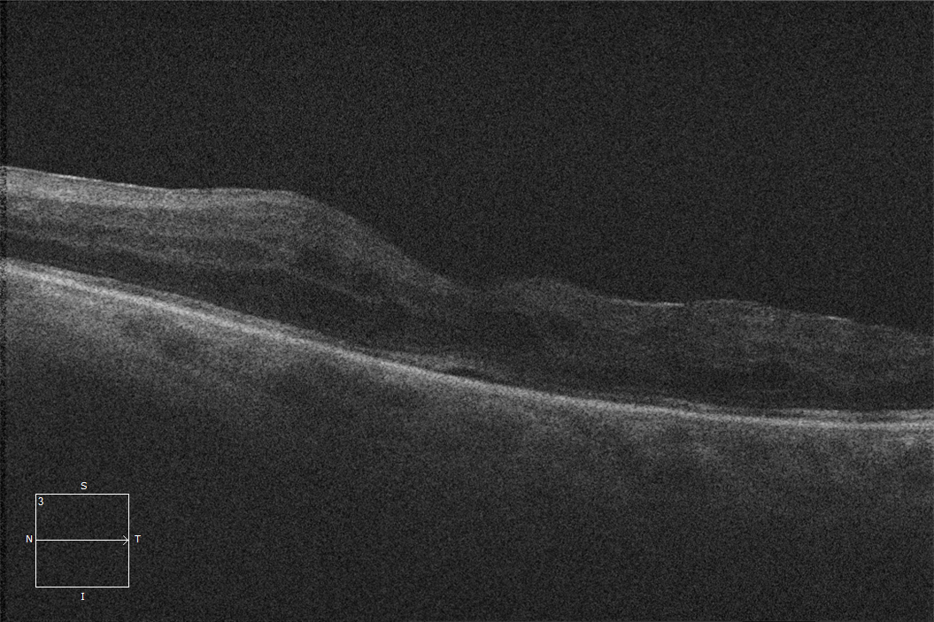
Dry Age-Related Macular Degeneration. 80-year-old woman with central geographic atrophy. The choroidal atrophy and variable drusen morphology are typical of AMD.
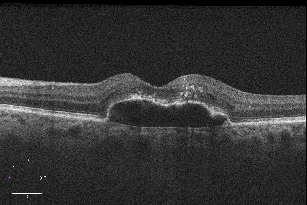
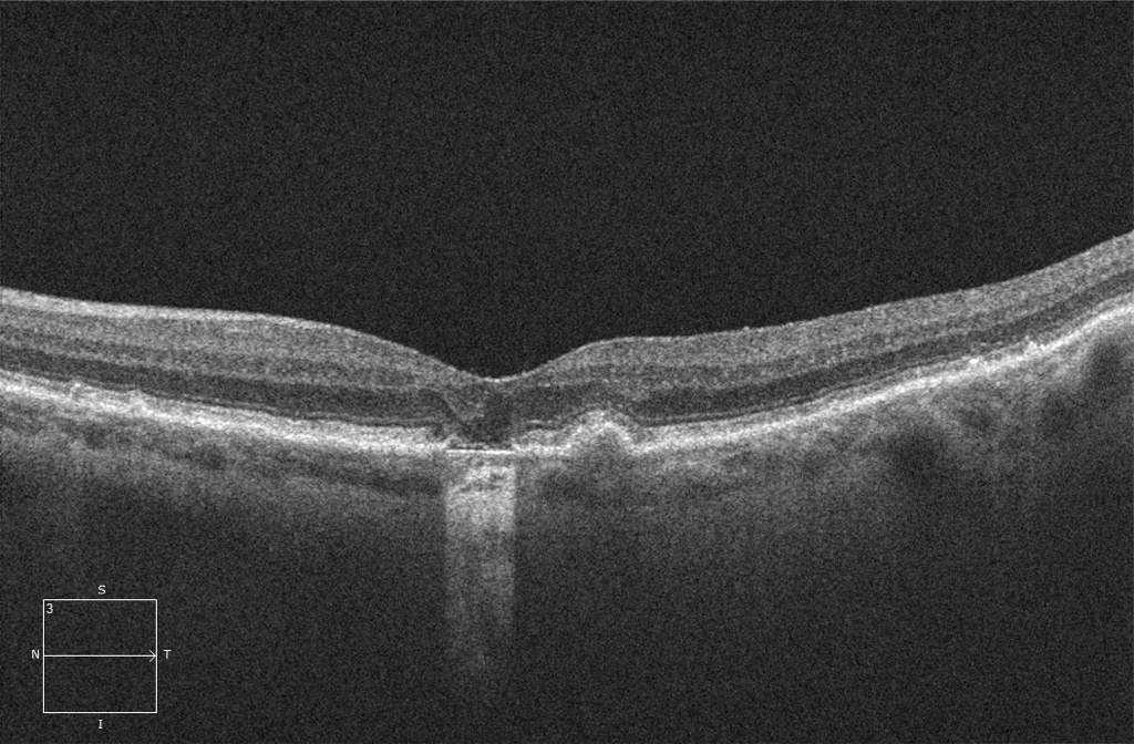
Lamellar Macular Hole. 55-year-old woman with an epiretinal membrane that has resulted in a lamellar hole configuration. The schisis is secondary to the membrane.
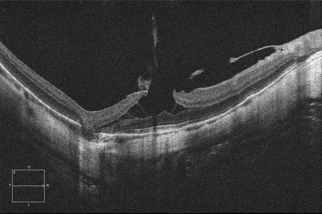
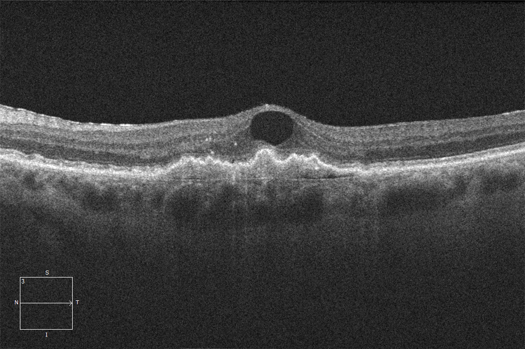
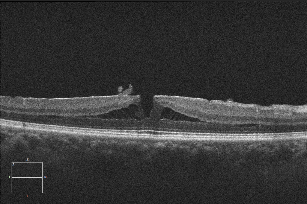
High Myopia plus Lamellar Macular Hole. 45-year-old woman with high myopia (-15 diopters). In such cases, macular schisis can be detected with or without an overlying epiretinal membrane.