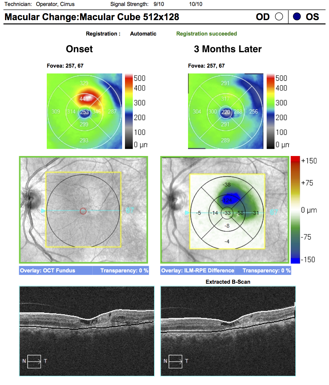
62-year-old woman with 3 monthly anti-VEGF injections. The images presented compare the presenting OCT to the follow-up OCT. The central cross sections do not demonstrate significant change. Despite the quantitative decrease in retinal thickness the qualitative changes are only detectable eccentric to the foveal center. Compare cuts 67 to cuts 46. In cuts 67 there appears to be little change (the cystoid changes are flat without internal speckling consistent with chronicity). However cuts 46 clearly demonstrate a decrease in subretinal hyperreflective material and resolution of the intraretinal fluid.