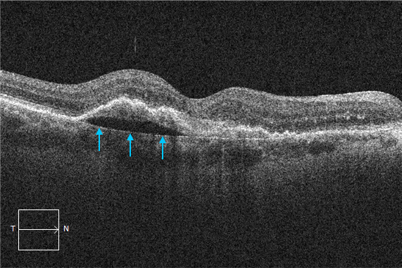
What is that line?
OCT interpretation relies on correctly identifying several key structures. One of these is the retinal pigment epithelium (RPE). However, in cases of pigment epithelial detachments, particularly in older patients, there is another line visible below the RPE.
This represents Bruch’s membrane.
It is important not to confuse it with the RPE or you may erroneously assume there is subretinal fluid.
Want to be able to interpret OCTs like an expert?
We can show you how!