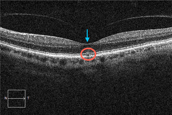
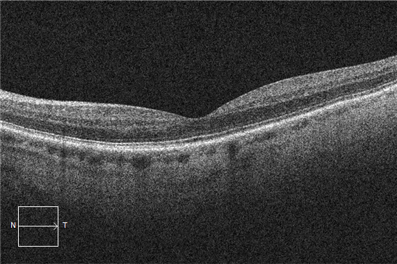
Full thickness macular hole formation commences in the outer retina and progresses inward. In this case there is vitreomacular adhesion (arrow) without obvious inner retinal distortion. However, it is still reasonable to classify this case as vitreomacular traction due to the outer retinal changes (circle). After 2 months the patient underwent spontaneous release of the posterior cortical vitreous (shifted beyond the view of this OCT image) from the retina and the outer retinal tissue normalized as a result. This is not a rare finding. Just remember to look for it on OCT testing.