From our May 2, 2016 newsletter.
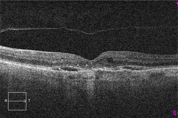
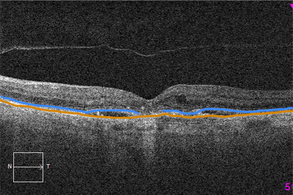
Distinguishing between the ellipsoid zone and the retinal pigment epithelium (RPE) is one of the most important steps when qualitatively evaluating an OCT. The ellipsoid zone is a consensus term based on current understanding of OCT lexicon and landmarks. It represents the inner portion of the photoreceptor layer that is densely packed with mitochondria. Meanwhile the RPE is a longstanding and well-described term referring to the cell layer that photoreceptors use for nourishment (among other roles). The reason this becomes important is that subretinal fluid will elevate the retina and a bright white band can be seen. But this is not a pigment epithelial detachment (PED). Once you are able to confidently distinguish the ellipsoid zone and the RPE, then you will be one step closer to becoming an expert at OCT interpretation.Tip
The RPE is labelled in light brown and the ellipsoid zone is labelled in light blue. Note the central geographic atrophy with resultant loss of the ellipsoid zone (discontinuous space).A Clear Vision for Professional Development
Elevate your eye care skills with image-rich content and relevant case examples that empower you to treat patients more effectively.
Send this Newsletter to a Friend
