Let’s talk about Retinal Arterial Occlusion (RAO), a vision-threatening pathology with visual outcomes ranging from 20/25 to no light perception. Even though we can often diagnosis RAO on clinical examination, adjunctive OCT imaging can still be useful. That’s because the OCT appearance is nearly pathognomonic and demonstrates thickening and inner retinal opacification in the acute phase. It’s often followed by atrophy as the situation progresses and neural tissue is lost.
While understanding these OCT findings is important, a recent publication by Chen et al took it a step further. The authors correlated the ratio of pixel intensity of the inner versus outer retinal layers on visual outcome. Basically, they found that a greater pixel intensity in the inner retina (and a lesser pixel intensity in the photoreceptor region) was correlated with a worse visual prognosis.
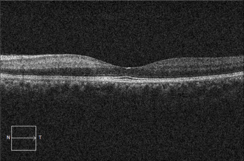
Figure 1: This OCT demonstrates a Branch RAO. Compare the area of high pixel reflectivity (nasal half) with the area of normal pixel reflectivity (temporal half).
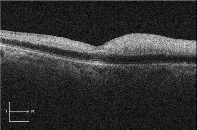
Figure 2: This OCT demonstrates a Central RAO with highly reflective internal pixelation. The patient went on to a poor outcome with 20/400 vision.
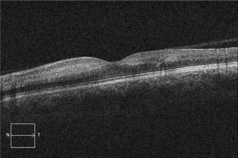
Figure 3: This OCT demonstrates a Central RAO with less reflective pixelation than seen in Figure 2. This patient went on to a modest outcome with 20/50 vision.
Let’s put this into perspective. For years, radiologists have been quantifying the pixel intensity to determine lesion characteristics on CT and MRI scans. But now, we have the first study doing the same for arterial occlusive disease of the retina. If this biomarker proves itself in larger studies, then we may be able to predict visual outcomes based on the presenting OCT!
In the meantime, keep reviewing your scans, as other disease processes also present on OCT with varying extents of hyperreflective pixelation (think cotton wool spots, some Retinal Vein Occlusions, hard exudates, etc). The authors actually used a neat trick to create a pixel ratio comparing the inner and outer retina. Maybe someday we’ll be using a “pixel ratio score” to confirm diagnoses and predict outcomes.
Always learning,
The EyeCarePD Team


