From our July 25, 2016 newsletter.
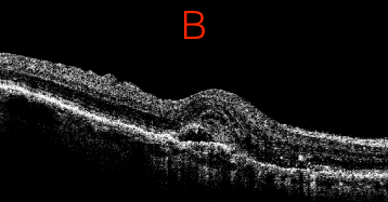
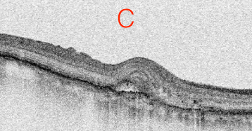
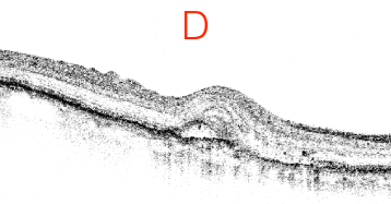
You may or may not have read the recent journal article that evaluated the utility of contrast alteration in macular OCT interpretation. Specifically, this study looked at whether macular OCT images viewed under different contrast and background color settings made a difference to the readers’ ability to detect specific structures and lesions. You can check out all the details here.Tip
When viewing macular OCT images, adjust the contrast and background color settings to help you see subtle differentiations. The results of the study show that contrast alteration can indeed be important in detecting various features. However, there’s no single setting that’s best for all images and features. So here’s what we recommend: In daily practice, keep it top of mind that image manipulation is not only possible, but important, since subtle differentiations can alter your diagnosis or treatment.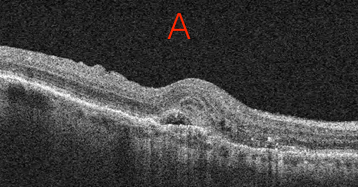
A Clear Vision for Professional Development
Elevate your eye care skills with image-rich content and relevant case examples that empower you to treat patients more effectively.
Send this Newsletter to a Friend