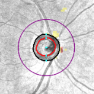
RNFL Deviation Map in a Normal Patient
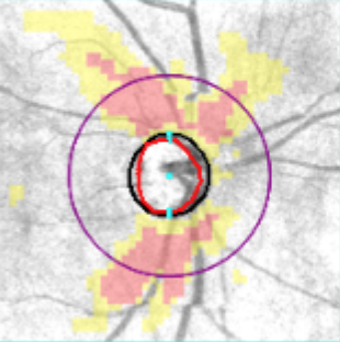
RNFL Deviation Map in a Glaucomatous Patient
How do you follow your patients who are Glaucoma Suspects? What about those with definitive Glaucoma? Does your algorithm vary by the extent of disease (mild, moderate, severe)?
In a seminal publication by Zhang et al on behalf of the Advanced Imaging For Glaucoma Study Group, the authors set out to answer the most basic and important of questions: What is the potential use of OCT, complementary to or in place of visual field (VF) testing, at varying stages of Glaucoma.
For years we have been using OCT-based nerve fiber layer (NFL) measurements. Then we added ganglion cell complex (GCC) analysis into our armamentarium. We have continued to use VF testing and incorporated everything into one big mix. Prior studies supported these clinical trends, but Zhang et al’s study provides tremendous evidence using disease cross-sectional analysis, thus enhancing our confidence in these biomarkers.
This publication by Zhang and colleagues is on my “Must-Read List” for all doctors, but let me sum it up with a few simplifications:
- Structural changes proceed functional changes in early disease. This means that if you have a patient who is a Glaucoma Suspect or has Preperimetric Disease, then you are better off using OCT analysis (RNFL/GCC) as opposed to VF testing to monitor for progression.
- In cases of early Glaucoma, RNFL and GCC analysis provide a higher detection rate for progression than VF (similar to the findings in Suspect and Preperimetric Disease).
- But, in cases of moderate or advanced Glaucoma, the VF is a more accurate biomarker than RNFL by OCT for detecting progression. However, GCC by OCT remains as effective as VF for detecting progression.
Does this mean that I am telling you to stop VF testing in early cases and to stop RNFL OCT testing in later cases? Of course not! The authors even note that cases will be missed if you do not combine OCT and VF data. After all, clinical care is more of an art than a science. But these rules will help solidify your management plan and care for your patients in the real world (where all the data are not perfect and patient cooperation and image quality can vary).
With this knowledge at the forefront of our attention, EyeCarePD is proud to announce our newest educational product, INTERPRET: Glaucoma. This FREE program provides hundreds of examples of OCT analysis and has a proven benefit to improve your OCT interpretation skills.
PLAY NOW. IT’S FREE!
Always learning,
Dr David Lederer and The EyeCarePD Team