With our recent post discussing the 2017 publication from the Advanced Imaging For Glaucoma Study Group, we thought now would be a good time to pick out a few of the more intriguing statements to bring to your attention. So let’s jump right in.
- Visual field (VF) testing is difficult for some patients. This is particularly true in those with advanced glaucoma.
- The retinal nerve fiber layer (RNFL) reaches a minimum thickness value and basically plateaus in advanced disease. This means that once the RNFL reaches the range of 45-65 microns (depending on your device), even if the disease advances, the RNFL will not change.
- Interestingly, the ganglion cell complex (GCC) will continue to decrease later in disease. This means that in more advanced cases, the best OCT parameter to follow is not the RNFL (as it will plateau), but rather the GCC.
- Be careful with signal strength. Signal strength standardization remains important across time, as small segmentation errors can lead to errors in the progressional analysis.
- Be careful with age. Age-related thinning of the RNFL and GCC is important to consider. And be certain that your OCT device software ensures age-matched controls not only in individual scan analysis, but also in progression analysis (which can vary by manufacturer).
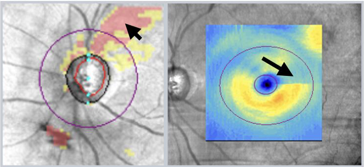
The RNFL deviation map demonstrating a superior arcuate defect (arrow). The same patient also demonstrates a superior defect in the ganglion cell complex (arrow).
OCT analysis of the RNFL is now commonplace. GCC analysis is gaining momentum and use. Progression analysis software continues to improve and progression can now be tracked for both the RNFL and GCC. VF testing remains critical to ensure that moderate and advanced cases are not missed and for guiding treatment decisions. By combining OCT analysis of the RNFL, GCC analysis and VF testing, our management of glaucoma continues to become more objective and refined.
Always learning,
Dr David Lederer and The EyeCarePD Team
Get the FREE OCT sample course!
