From our July 4, 2016 newsletter.
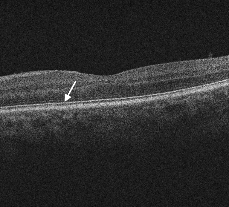
When you look at an OCT on your computer screen to evaluate it, you’re actually seeing many raw data points grouped together. Each point is detected by the OCT device, and then converted to a pixel and displayed at a specific intensity. The OCT device can detect thousands of levels of intensity (ranging from 4,000 to more than 65,000), but your computer monitor usually only displays 256 levels of intensity. You may be thinking, “interesting… but so what?” Well, this simple fact explains a common problem in OCT image interpretation; if you look at an OCT in pseudocolor, you’ll notice many artificial transition points between each pixel, which can lead to false interpretations.Tip: Help prevent misinterpretations by viewing OCT images in grayscale.
In the past several years, experts have been moving away from pseudocolor and opting for grayscale images. This has been proven in the OCT literature as being particularly important in preventing misinterpretations of subtle ellipsoid zone, retinal pigment epithelium and epiretinal membrane findings.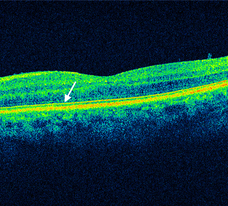
A Clear Vision for Professional Development
Elevate your eye care skills with image-rich content and relevant case examples that empower you to treat patients more effectively.
Send this Newsletter to a Friend