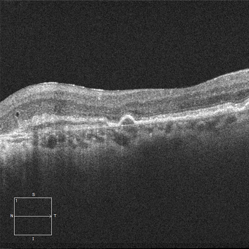
ORT stands for outer retinal tubulation. It is theorized to be due to degenerated photoreceptors that become arranged in a circular or ovoid pattern.
A Clear Vision for Professional Development

ORT stands for outer retinal tubulation. It is theorized to be due to degenerated photoreceptors that become arranged in a circular or ovoid pattern.
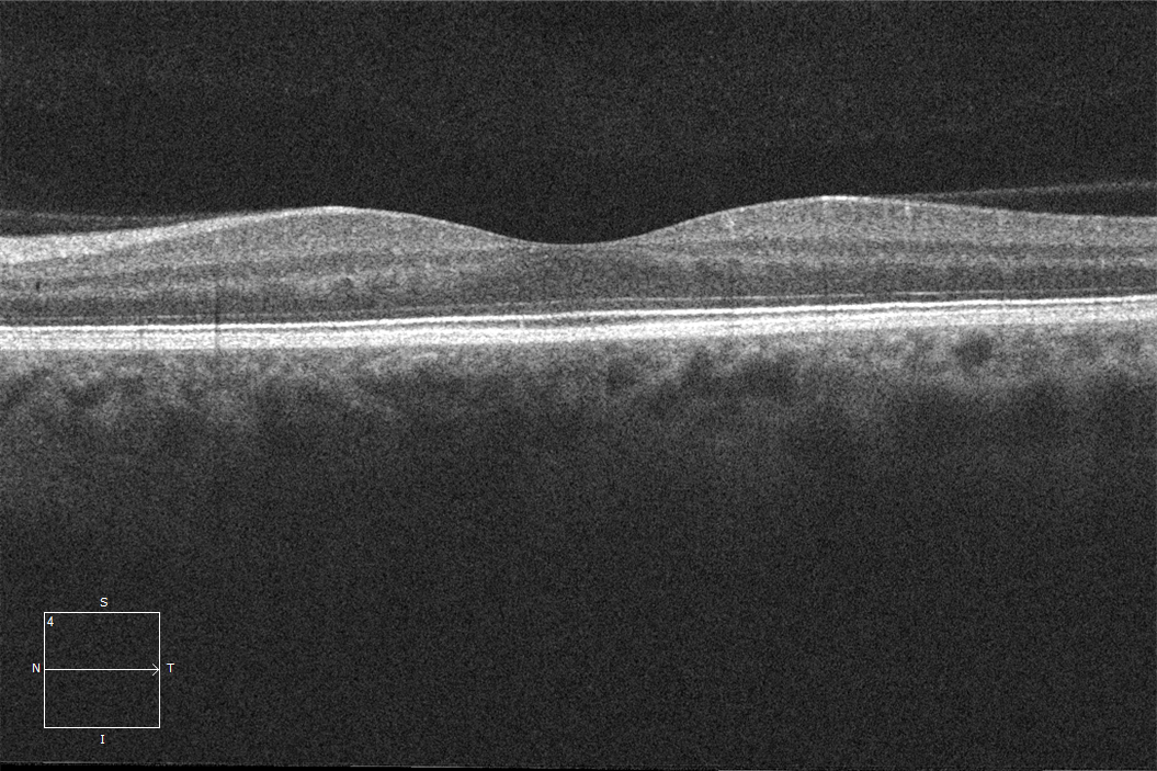
Inner retinal opacification is a common finding seen on OCTs for retinal artery occlusions.
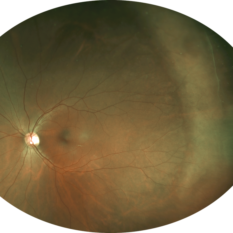
The image displayed is not a diffuse amelanotic choroidal melanoma.
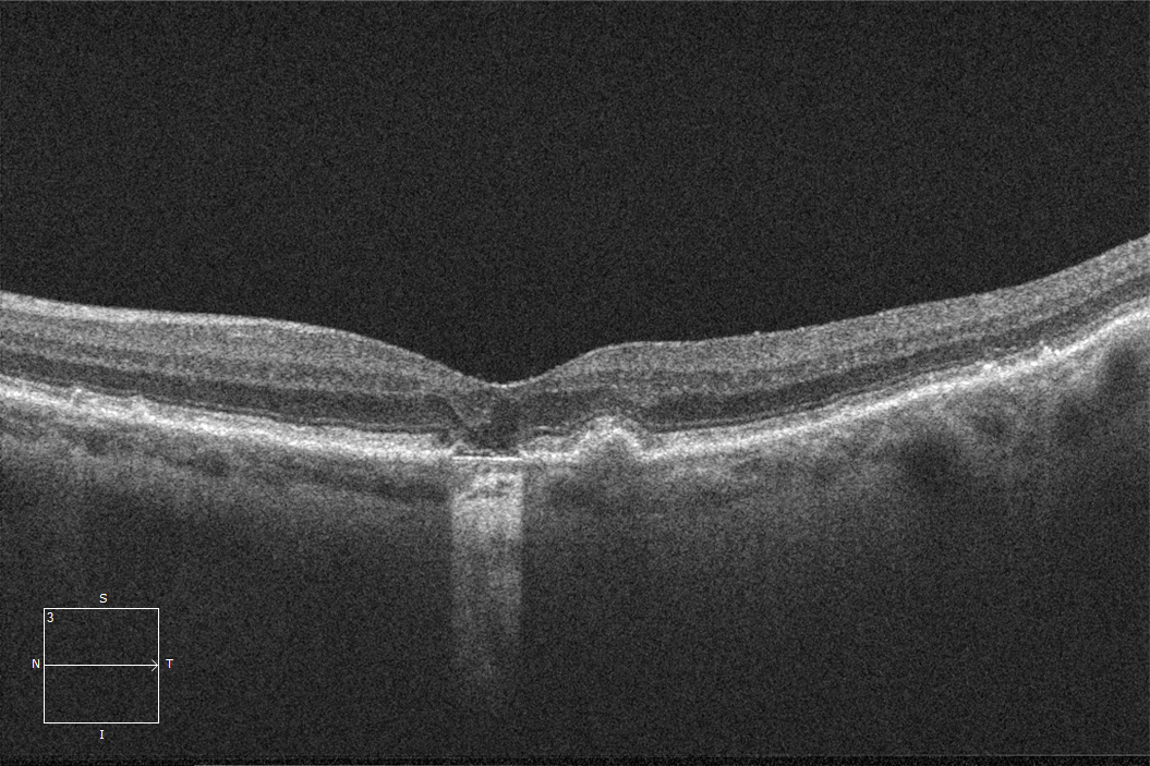
This OCT image is typical of dry age-related macular degeneration complicated by focal atrophy.
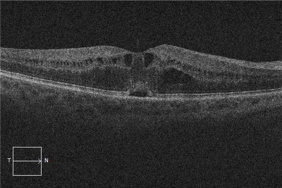
Let’s continue the conversation about fluid. But this time, the spotlight is on intraretinal fluid.
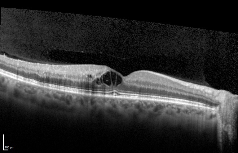
This OCT image is typical of cystoid macular edema after a retinal vein occlusion.
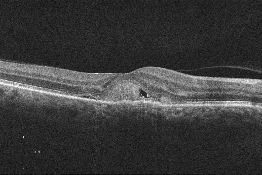
This OCT image is typical of choroidal neovascularization.
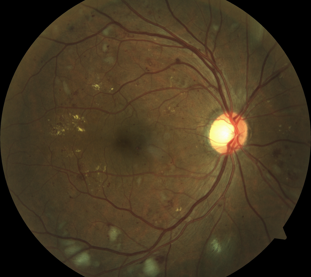
43-year-old man with no known past medical history presents for a routine eye exam.
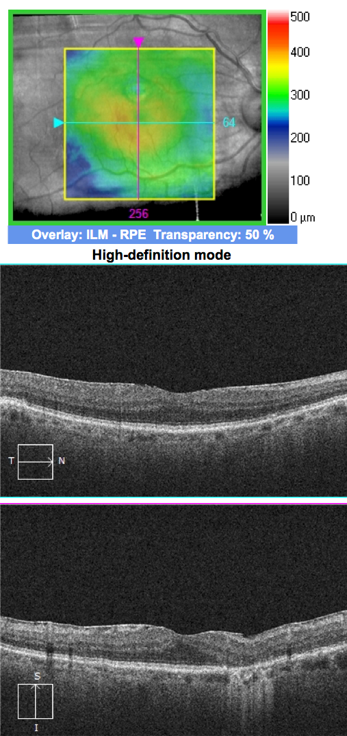
83-year-old man with Dry Age Related Macular Degeneration and an Epiretinal Membrane.
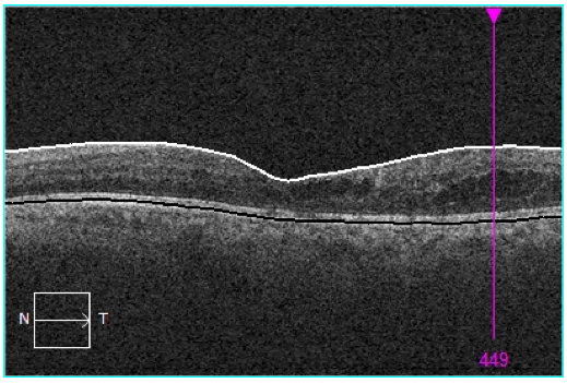
73-year-old man with diabetic retinopathy and prior panretinal photocoagulation for proliferative disease that is now regressed and stable.
EyeCarePD Inc.
All Rights Reserved
By using this site you agree
to our Terms and Conditions.